New technology puts a spatial lens on CRISPR screening

Recently, scientists have been able to explore gene circuitry in individual cells using methods that suppress particular genes and measure the impact on the expression of other genes. These methods, however, fail to capture spatial information such as the effects from, or on, neighboring cells, which can provide important clues to a cell or gene’s role in health and disease.

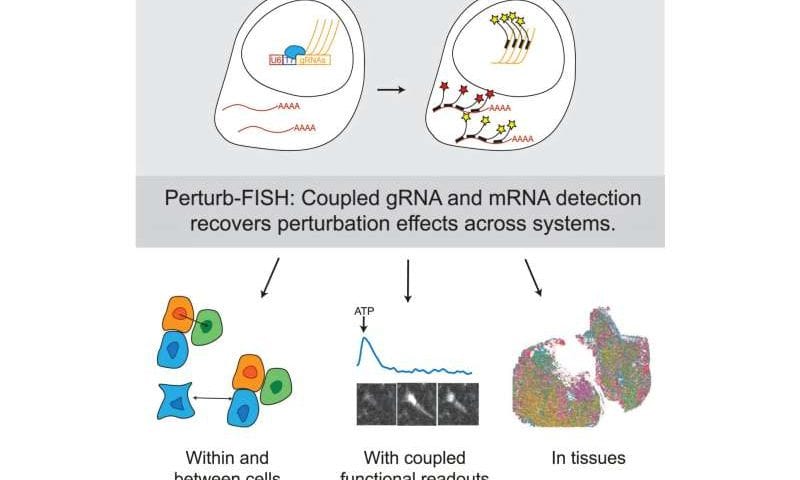
Now, a technology developed in the Spatial Technology Platform at the Broad Institute of MIT and Harvard builds on these methods with cutting-edge spatial advances. Their method, known as Perturb-FISH, combines imaging-based spatial transcriptomic measurements with large-scale detection of CRISPR guide RNAs.

The researchers demonstrated Perturb-seq’s ability to uncover new cellular and functional insights, including the effects of autism-related genes on cellular activity and the interactions between human tumor and immune cells in an animal model. Further refinements to Perturb-FISH could make it even more widely accessible and enable a range of new biological investigations. The technology and demonstrations of its utility are published in Cell.

“With Perturb-FISH, we’ve developed a powerful new way to examine the roles of genes and genetic circuits in tissue development, homeostasis, and dysfunction,” said co-senior author Sami Farhi, director of the Spatial Technology Platform at the Broad Institute.

“Our team is dedicated to devising spatial tools for the benefit of the scientific community, and we hope this new method is just the first of many more that we’ll build and share.” Farhi led the work along with co-senior author Brian Cleary, a former Broad Fellow and Merkin Institute Fellow who is now an assistant professor at Boston University.

Gone FISH-ing

Previously, researchers had combined single-cell RNA sequencing with CRISPR screening to examine gene networks within cells using tools such as Perturb-seq, but the cells’ spatial context wasn’t captured. Another method known as optical pooled screening measures the effects of gene-editing perturbations on cell fitness or other phenotypes, but doesn’t capture gene transcriptional states.

Scientists in the Spatial Technology Platform aimed to develop a comprehensive method that could measure at once which genes are altered in a cell, which genetic perturbation caused the changes, and the location of those affected cells in relation to other cells.

Key developments that made Perturb-FISH possible were computational methods developed by Cleary and a new way of amplifying the signal from single molecules (either CRISPR guide RNAs or gene transcripts) so they can be detected over background levels of fluorescence.

Traditional amplification methods don’t work with molecules as small as guide RNAs, so first author and former postdoctoral researcher Loϊc Binan devised an innovative strategy to generate many local copies of each guide RNA at its original site. By combining that with a fluorescence-based spatial transcriptomic method called MERFISH, Perturb-FISH can reveal both the identity of each perturbation and the cell’s transcriptome in their spatial context.

Genome editing technique enables partial inhibition of gene function in crops

Researchers at Kumamoto University have successfully executed a practical trial of a new genome-editing technique that allows for the partial inhibition of essential gene function without causing lethality. This innovative approach, which leverages CRISPR-Cas9 technology, opens new doors for genetic research and agricultural advancements by enabling the study and modification of genes that were previously difficult to analyze.

Genes that are crucial for survival often pose a challenge for researchers, as completely disabling them can be fatal to the organism. Traditional gene knockout methods, including CRISPR-Cas9, typically result in complete loss of function, making it difficult to investigate these vital genetic components.

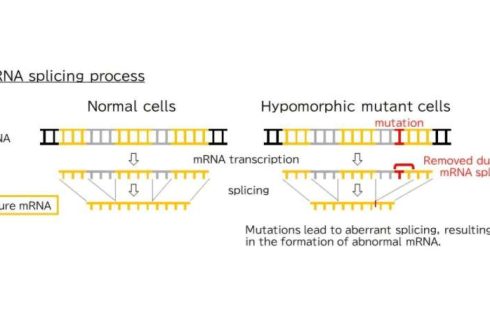
To address this limitation, the research team led by Associate Professor Takashi Ishida, Faculty of Advanced Science and Technology, Kumamoto University, conceived a method to introduce hypomorphic mutations, which reduce—but do not entirely eliminate—gene function.

Using the model plant Arabidopsis thaliana, the team successfully applied their method to the HPY2 gene, which is essential for cell division and plant growth. While complete loss of this gene results in seedling lethality, the newly engineered hypomorphic mutants exhibited delayed growth but remained viable, paving the way for researchers to analyze gene function in greater detail in future studies.

Applications for agriculture and beyond

This breakthrough, published in the Journal of Plant Research, has significant implications beyond basic genetic research. By fine-tuning gene activity instead of completely disabling genes, this technique could be used to enhance desirable traits in crops without causing harmful side effects. For example, modifying genes responsible for stress resistance in plants could lead to crops that are more resilient to climate change while maintaining their growth and yield.

“This method provides a powerful tool for studying genes that were previously inaccessible due to their essential nature,” said Associate Professor Ishida. “It also opens up new possibilities for precision breeding in agriculture, where controlled gene modification is key to improving traits without unintended consequences.”

DNA barcodes enable high throughput RNA and protein detection in deep tissue

For the Liu Lab, necessity is truly the mother of invention. The researchers were examining how the 3D organization of the genome controls development and needed to image hundreds of RNA molecules in a thick tissue sample to understand where and how genes were being expressed in cells. There was just one problem: There weren’t any tools that were up to the task.

One technique could image lots of RNA molecules, but only in a thin layer of cells. Another method could image in deep tissue but could only detect three or four molecules in a single sample.

So, the team at HHMI’s Janelia Research Campus decided to build their own tool. The result is an innovative new technique that uses a novel DNA barcode system to track hundreds of RNA and protein molecules in single cells within thick biological samples, providing researchers with a full picture of how these structures are organized inside tissues. The research is published in the journal Science.

RNA molecules carry instructions from DNA for making proteins that carry out much of the work of living cells. Knowing where these molecules are located in complex tissues is a critical part of understanding where and how genes are being expressed across different regions and cell types. This information enables researchers to decipher how genes function in different parts of an organism, how they enable development, and how they might be altered in diseases.

Beyond its use in biology and neuroscience, the new method could also potentially be used in diagnostic imaging, according to the researchers.

“I think it will be a gamechanger very broadly, not just for people in my field,” says Janelia Group Leader James Liu. “It was a tool developed to answer a very obscure question, but I think all biologists can use the technique in their favorite samples.”

Depth and throughput

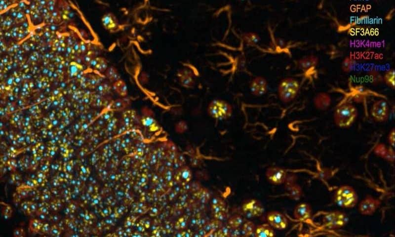
The new imaging tool, called cycleHCR, builds on a previously developed technique called Hybridization Chain Reaction, or HCR. The technique assembles multiple fluorophores on a target that shine like a bright beacon when imaged by a fluorescence microscope, enabling researchers to see molecules in single cells deep inside tissues.

But HCR is limited by current fluorophores, which, because of the wide spectrum they cover, only allow three or four colors to be used at one time. This means researchers can only detect a handful of molecules in a sample, making it difficult to get a full picture of how they are organized within tissues.

To overcome this, the team designed novel DNA barcodes that they could attach to the targets. Just like barcodes on products in a supermarket, which designate every individual type of item in the store, the unique DNA barcodes allow the researchers to tag each type of molecule in the sample.

Each barcode contains two parts. When the two pieces match, the target is amplified by the HCR technique. The two parts of the barcode ensure the tags are specific enough to detect individual types of RNA molecules.

The barcodes were also designed to be easily removed, so multiple rounds of HCR can be performed on the same sample. The initial round of imaging uses three barcodes, picking up three RNA molecules in three different colors. These barcodes are removed, and a second round of imaging uses three different barcodes and so on for multiple rounds, eventually allowing the discovery of an unlimited number of targets in a single sample.

“We modified the split amplification chain reaction technique in a way that now we are adding barcoding to it where we can detect hundreds, potentially even thousands of RNAs, with these multi rounds,” says Valentina Gandin, a Senior Scientist in the Liu Lab who co-led the research. “The barcoding was a novelty that we added to this.”

In addition to detecting RNA, the researchers developed a way to use the same barcodes to detect proteins, allowing researchers to better understand how both RNA and proteins are organized in tissues.

The team also automated the system, enabling the researchers to detect up to a dozen molecular species in a single day without having to constantly monitor the process. Additionally, Postdoctoral Scientist Jun Kim, a co-first author of the new study, developed analysis methods that map where genes are expressed spatially and help researchers make sense of the raw data. The Liu Lab also worked with the Scientific Computing Software support team at Janelia and developed an automated pipeline for processing the large amount of imaging data generated.

The researchers worked with other labs at Janelia to use the new method to quantify gene expression in mouse embryos, quantifying 254 genes in a single sample. This enabled the researchers to characterize all the cell types in the embryo and discover new cell types that had not been previously characterized, information that is important for understanding development.

The new technique has already generated attention in the scientific community, and the team is working to enable more labs to use the new tool. They shared all of the barcode sequences so other labs can design their own probes, even if they don’t have an automated system. The team is also building a more streamlined version of their prototype system and plan to share their automated platform with the scientific community.

“Eventually we want everybody to use it,” Liu says. “We would really like our technique to be broadly spread to enable every scientist to be able to use it.”

Engineered cell cultures boost anti-inflammatory compounds in endangered snow lotus

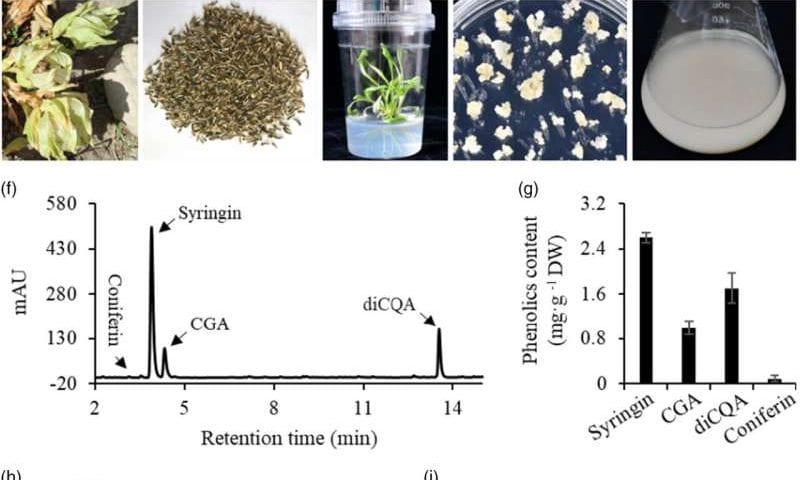
Researchers from the Qingdao Institute of Bioenergy and Bioprocess Technology (QIBEBT) of the Chinese Academy of Sciences (CAS) have developed a novel method to enhance the production of valuable medicinal compounds in the endangered snow lotus (Saussurea involucrata). Their study is published in the Plant Biotechnology Journal.

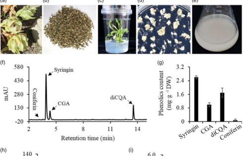
The researchers achieved a 15-fold increase in syringin, a bioactive compound with potent anti-inflammatory properties, by genetically engineering suspension cell cultures. This advancement holds promise for scalable applications in pharmaceuticals and functional foods.

The snow lotus, native to China’s Tianshan Mountains, has long been prized in traditional medicine for its ability to treat conditions such as rheumatoid arthritis and high-altitude sickness. However, its slow growth and vulnerability to environmental stressors have pushed the species to the brink of extinction. To address this, the researchers turned to suspension cell cultures, which can be rapidly scaled in bioreactors, bypassing the challenges of traditional cultivation.

Using methyl jasmonate (MeJA) to stimulate syringin synthesis, the researchers conducted transcriptomic analysis and identified two key glycosyltransferase genes, SiUGT72BZ2 and SiUGT72CY1. Overexpression of SiUGT72BZ2 in suspension cell cultures resulted in syringin levels 15.2 times higher than those in control groups, alongside a significant increase in coniferin, another anti-inflammatory compound. In contrast, overexpression of SiUGT72CY1 yielded only a 5.9-fold increase in syringin.

When tested on lipopolysaccharide (LPS)-induced inflammatory cells, extracts from SiUGT72BZ2-enhanced cultures demonstrated a 10.6% greater suppression of interleukin-6 (IL-6) production compared to controls at lower doses. “The synergistic effects of syringin and coniferin likely contribute to this enhanced anti-inflammatory activity,” explained co-author Dr. Wu Zhenying.

“This system decouples compound production from environmental constraints, making it a viable solution for large-scale production,” said Ph.D. candidate Xu Yue, a co-first author of the study.

By leveraging genetic engineering and suspension cell culture technology, the researchers have opened new avenues for conserving the endangered snow lotus while meeting the growing demand for its medicinal compounds. This study underscores the potential of biotechnology to address both conservation and industrial challenges in producing plant-based therapeutics.

Roche Genentech Innovation Center Boston Launched at Harvard’s Enterprise Research Campus

The Roche Genentech Innovation Center Boston has been launched at Harvard’s Enterprise Research Campus in Allston, MA. Roche officials say the move will further strengthen the partnership in areas such as disease biology, engineering, and artificial intelligence/machine learning.

The Center will serve as Roche’s Cardiovascular, Renal, and Metabolism (CVRM) hub that will also support some of Roche’s and Genentech’s efforts in AI and data science to accelerate drug discovery and development. The labs will be equipped for advanced research, and over time the center could employ up to 500 people, reports Roche. Manu Chakravarthy, MD, PhD, senior vp and global head of CVRM Product Development, will be the center’s site head.

With the new center, Roche and Genentech are expanding their presence within the Greater Boston innovation ecosystem, leveraging local talent and anchoring an entrepreneurial hub at Harvard’s Enterprise Research Campus, according to a Roche spokesperson, who adds that this new investment highlights Roche’s dedication to advancing healthcare through academic and scientific collaboration.

The hub has also been created to strengthen an existing relationship between Harvard and Roche that has been in place for over a decade. These drug discovery efforts have included work to combat antibiotic resistant bacteria as well as the use of AI in cancer research to identify targeted approaches to treatment and to analyze the effectiveness of new cancer drugs in clinical trials.

“Roche has a significant presence in the U.S., and this new center is one of a number of research and manufacturing investments that Roche is making in the U.S. through Genentech and Roche Diagnostics,” says Thomas Schinecker, CEO of Roche.

“In a region grounded in innovation in the sciences and health, when leading biomedical companies and research universities work together, breakthroughs follow,” adds Alan Garber, president of Harvard. “We are delighted that the Roche Genentech Innovation Center will launch the first phase of the Enterprise Research Campus. The new Center will bring new talent and new opportunities to pursue our shared commitment to discovery for the advancement of human health.”

Value chain in the United States

Roche has the entire pharma and diagnostics value chain in the United States with four Genentech sites spanning R&D, production, and distribution in its pharmaceutical division and seven sites focused on R&D and manufacturing in Diagnostics, points out Schinecker, who notes that the company has more than 25,000 employees in America, invested $11 billion in the United States in the last 10 years, and will continue investing in the country.

Harvard’s Enterprise Research Campus, the first phase of which is underway and developed by Tishman Speyer and Breakthrough Properties, has been intentionally designed to foster innovation and collaboration. Located adjacent to Harvard’s Science and Engineering Complex and near Harvard Business School, the ERC will be a mixed-use development integrating research and laboratory space, residential buildings, a hotel, a conference center, retail units, and a variety of community-centered outdoor spaces. The first phase of construction is expected to be completed in 2026.

Sex Hormone Protects Against Opioid Misuse in Female Rats

Most people who misuse opioids take the drugs to relieve pain, but data suggest that men are more likely to misuse and overdose on opioids than women, even though they suffer less chronic pain. Scientists hypothesize that something other than, or in addition to, chronic pain must be putting men at higher risk of developing problems managing their opioid use.

Newly reported research in rats by scientists at Washington University School of Medicine in St. Louis suggests that one underlying cause may be biological. The study showed that male rats in chronic pain gave themselves increasing doses of an opioid—specifically, fentanyl—over time, while female rats with the same pain condition kept their intake at a constant level, similar to what is seen in people. The behavioral difference was driven by sex hormones, the researchers found. Further experiments showed that treating male rats with the hormone estradiol (E2) led the animals to maintain a steady level of fentanyl intake.

The study findings indicate that differences in how men and women use and misuse opioids may be driven by their hormones, and that a deeper understanding of how sex hormones interact with chronic pain could open up new approaches to addressing the opioid epidemic.

“These data suggest that men may be inherently predisposed to misuse opioids in the context of pain because of their balance of sex hormones,” said Jessica Higginbotham, PhD, a postdoctoral researcher in the lab of Jose Moron-Concepcion, PhD, the Henry Elliot Mallinckrodt Professor of Anesthesiology at WashU Medicine.

First author Higginbotham, senior author Moron-Concepcion, and colleagues reported on their findings in Neuron, in a paper titled, “Estradiol protects against pain-facilitated fentanyl use via suppression of opioid-evoked dopamine activity in males.” In their paper, the team concluded, “While pain relief is a primary motivator for opioid misuse, our findings highlight a critical role for hormonal regulation in opioid vulnerability under conditions of pain and begin to address a major gap in our current understanding … This study reveals nuanced crosstalk between hormones and pain states that may alter reward pathway dysfunction perpetuating opioid use.”

Opioids are potent analgesics used to treat the symptoms of pain, but they are highly prone to abuse, the authors wrote. And while pain relief is the most frequently reported motivation for opioid misuse, the team stated, “Despite sex and gender differences in pain and opioid use—with women reporting greater pain, while men are more frequently misusing opioids and overdosing—the underlying mechanisms remain unclear.”

When a person—or a rat—takes an opioid such as fentanyl, it acts on the brain in two ways. The drug blocks the transmission of pain signals, relieving pain, and it triggers the release of dopamine from the reward center in the brain, creating a feeling of euphoria. Previous work by Moron-Concepcion and members of his laboratory had shown that pain itself affects dopamine levels in the brain, suggesting that opioids and pain may interact to influence dopamine levels. “The transition to compulsive drug use stems from neuroadaptations within the mesolimbic dopamine (DA) circuitry, a key regulator of reward and motivation,” the team continued. “Opioid reinforcement triggers extracellular DA release in the nucleus accumbens (NAc) through disinhibition of ventral tegmental area DA (VTADA) neurons.”

To understand how pain influences opioid-seeking behavior in sex-specific ways, Higginbotham and Moron-Concepcion studied rats with induced chronic pain in their paws. The researchers found no difference between males and females in terms of how much pain the animals experienced, as measured by how quickly they drew back their paws when touched. They also found no difference between the sexes in how much pain relief a dose of fentanyl provided, using the same measurement. But the team found that males allowed self-administration (SA) of fentanyl went back for more and more of the opioid over the course of the three-week study, while the females did not. “Using a model of persistent inflammatory pain, intravenous (i.v.) fentanyl self-administration (SA) and wireless in vivo fiber photometry, we identify sex-specific mechanisms underlying maladaptive opioid use,” they explained in their paper.

The researchers discovered an important difference between male and female rats in the amount of dopamine released after a dose of fentanyl. Females produced the same amount of dopamine from fentanyl over the course of the experiment, regardless of whether they were in pain or not. Males that were not in pain responded like females. In contrast, males in chronic pain generated a bigger and bigger dopamine response to fentanyl over time. In other words, pain made the feel-good part of opioids feel even better for males, but not for females.

“We had thought that maybe the males developed a tolerance to fentanyl and needed increasing amounts to relieve the pain, but that wasn’t it,” said Moron-Concepcion, also a professor of psychiatry and neuroscience. “The males were taking more and more fentanyl to feel that ever-increasing high. In males, but not females, the pain condition itself affected the reward centers of the brain and drove them to take more drugs.”

Further experiments revealed that sex hormones were responsible for the different dopamine responses in male and female rats. Ovaries are the primary source of sex hormones in females, producing estrogen, progesterone, and small amounts of testosterone. The researchers found that female rats whose ovaries had been removed—ovariectomized (OVX) animals—responded to fentanyl similarly to males, with increasing amounts of dopamine released and an increase in opioid-seeking behavior. In contrast, males that were given estrogen (estrodiol, E2) exhibited dopamine responses and opioid-seeking behavior similar to that of females.

“By linking instrumental opioid use with real-time reward circuit activity across several weeks, we identify hormonally regulated pain-induced neuroadaptations within VTADA systems under hormonal regulation, which drive excessive opioid use,” the authors reported. “While pain relief is a primary motivator for opioid misuse, our findings highlight a critical role for hormonal regulation in opioid vulnerability under conditions of pain and begin to address a major gap in our current understanding.”

The findings suggest that a drop in estrogen levels with menopause may help explain why older women have higher rates of opioid abuse compared to younger women, Higginbotham said. “Our findings suggest that high E2—relative to other circulating hormones—may add resilience against opioid misuse in pain,” the team noted.

Moron-Concepcion stated, “What we can do now is start thinking about how to find the right balance of hormones to prevent opioid use disorder in people with chronic pain. We haven’t yet looked at the role of other sex hormones such as testosterone or progesterone. Is there a perfect combination of hormones that can reverse the effects of pain on opioid use? That’s something we’d like to find out.”

Higginbotham added, “We focused on estrogen in this study, but I doubt the effect we saw is due to estrogen alone. It is more likely to be the balance of all the sex hormones in the body that influences risk. Men and women have the same sex hormones, just in different amounts, and our data suggest that females have a more protective balance than males. But if that balance changes, the risk of developing opioid use disorder could change, too.”

Reporting on their findings, the authors further stated, “To advance pain treatment in the face of the opioid crisis, it is imperative that we delineate how biological sex and hormone mechanisms predispose certain populations for higher misuse liability and that we use this knowledge to improve individualized treatment strategies.”

Motion Sickness Brain Circuit Linked to Obesity in Mice

Motion sickness is a very common condition that affects about 1 in 3 people, but the brain circuits involved are largely unknown. Newly reported work outlines a brain circuit involved in motion sickness that also contributes to regulating body temperature and metabolic balance. Findings from the team’s work, including studies in a mouse model of motion sickness, may also point to new strategies for treating obesity.

Co-corresponding author, Yong Xu, MD, PhD, professor of pediatrics–nutrition and associate director for basic sciences at the USDA/ARS Children’s Nutrition Research Center at Baylor College of Medicine, and colleagues, reported on their studies in Nature Metabolism. In their paper, titled “Vestibular neurons link motion sickness, behavioral thermoregulation, and metabolic balance in mice,” the team concluded that their collective findings “… could help to provide potential targets to treat motion sickness in humans, and highlight the underappreciated function of the vestibular system in obesity control.”

Symptoms of motion sickness in humans, including facial pallor, nausea, and vomiting, have been well-documented and investigated, the authors wrote. Interestingly the researchers stated, “… research suggests a potential link between obesity and impairments in balance and sensory processing, which contribute to motion sickness.” In addition, they commented, “… many genetic variants associated with motion sickness are implicated in the regulation of glucose tolerance, insulin sensitivity, and body-weight balance.” However, the exact relationship between motion sickness, metabolic regulations, and the underlying mechanisms isn’t known.

The Xu lab works with mouse models to investigate how the brain regulates metabolism and how this may be related to obesity and inform the development of more effective obesity drugs. Mouse models offer an abundance of molecular and genetic tools, as well as relevant behavioral assays to elucidate the neural mechanisms underlying physiological responses.

Motion sickness hadn’t been a key focus for the lab’s research. “When Longlong Tu, PhD, a postdoctoral fellow in my lab, proposed to investigate the brain circuits involved in motion sickness, a condition for which he is highly susceptible, I was not very excited about the idea because it’s not one of the main interests of my lab,” said Xu. “However, I became more interested and supported Tu’s idea when he explained the emerging evidence suggesting a link between motion sickness and metabolic balance, which is one of my research interests.”

But there was a challenge—mice are incapable of vomiting, one of the main manifestations of motion sickness in people. But mice and humans subjected to motion sickness stimuli, such as experiencing horizontal motion back and forth for some time, do both demonstrate hypothermia, a reduction in body temperature. “Notably, motion sickness leads to changes in thermoregulation, as evidenced by a reduction in body temperature observed in mouse, shrews, rats, and humans,” the authors wrote.” Xu added, “This allowed us to develop a mouse model of motion sickness in which we measured core body temperature, physical activity, and brain activity as the animals experienced motion stimuli.”

The authors further explained, that this mouse model of motion sickness “… exhibits prominent hypothermia during provocation, accompanied by associated perturbed thermoregulatory and other physiological adaptations typical of what is seen in people experiencing the condition.”

Through their reported studies the researchers found that motion activates glutamatergic neurons—neurons that produce glutamate, the primary excitatory neurotransmitter in the central nervous system—in the medial vestibular nucleus parvocellular part (MVePC) of the brain. Activation of these MVePCGlu neurons is required and sufficient to mediate motion-induced thermal adaptations, they found. “We show that motion-activated neurons in the MVePC are glutamatergic (MVePCGlu), and that optogenetic stimulation of MVePCGlu neurons mimics motion-induced hypothermia by signaling to the lateral parabrachial nucleus (LPBN),” they further explained in their paper. The researchers validated the model by showing that motion sickness-induced hypothermia does not occur when the mice are given the anti-nausea drug scopolamine.

“Given that the activation of the MVePC→LPBN circuit mimics motion-induced hypothermia, we next investigated whether inhibiting this circuit would attenuate it,” the investigators wrote. Xu added, “We further studied this motion sickness circuit by inhibiting the MVePCGlu neurons in the absence of motion stimuli. Inhibiting these neurons led to an increase in body temperature, along with increased physical activity. These physiological alterations suggest that chronic inhibition of MVePCGlu neurons may result in a higher energy expenditure in mice.”

The authors further noted, “Chemogenetic inhibition of MVePCGlu neurons can almost fully abolish motion-induced hypothermia, as well as cold-seeking behavior. Moreover, inhibition of the MVePC→LPBN circuit largely alleviates provocation-induced hypothermic responses and reverses cold-seeking behavior.”

When the researchers investigated the potential metabolic benefits of chronic inhibition of MVePCGlu neurons they found that while the mice ate more they gained less weight and exhibited better glucose tolerance and enhanced insulin sensitivity, physiological responses associated with better health. The collective results, they noted, “… highlight that chronic inhibition of MVePCGlu neurons prevents diet-induced obesity through increased energy expenditure in female mice.” Xu stated, “These results highlight the underappreciated function of the brain’s vestibular system in metabolic balance, and further raise the possibility that better understanding of the neural basis for thermoregulation during motion sickness may provide unconventional targets for the treatment of obesity.”

And for first author Tu, the findings offer hope that a better understanding of the brain circuit for motion sickness could also lead to improved medications for his condition. “Overall, these findings highlight MVePCGlu neurons as a potential target for motion-sickness treatment and obesity control,” the authors concluded.

StockWatch: Trump’s FDA Nominee Eyes Rare Disease-Specific Review Pathways

Testifying before the Senate Health, Education, Labor, and Pensions (HELP) Committee on Thursday, President Donald Trump’s nominee for FDA Commissioner Martin A. Makary, MD, sought to assure senators that if confirmed, he would support efforts to expedite reviews of new drugs indicted for rare diseases, as well as tailor to individual diseases the agency’s regulatory review-to-approval pathways designed to advance drugs to treat them.

“We have to customize the regulatory process to the condition that we’re trying to be able to offer hope, so, if a condition affects 19 people in the world as a partial triplication chromosome 15 disorder does, or a disease that affects 52 kids in the world, we cannot require two randomized control trials,” Makary said, responding to a question from Sen. Lisa Murkowski (R-AK), according to a transcript released by her office. “We have to customize the regulatory process to what we’re trying to do if our goal is to try to provide safe and effective therapies.”

Makary, a surgeon and public policy researcher at Johns Hopkins University, was nominated “to course-correct and refocus the agency,” Trump said last November in announcing his choice to helm the FDA. Should he be confirmed, Makary would report to Health and Human Services (HHS) Secretary Robert F. Kennedy Jr., who has targeted the FDA for drastic changes on a host of biopharma and broader healthcare concerns as part of his commitment to “make America healthy again” by “go(ing) wild on health.”

“If confirmed, I hope to ensure the FDA holds to the gold standard of trusted science, transparency, and common sense to rebuild public trust and make America healthy again,” Makary said in his opening statement.

Murkowski noted that Makary has committed to pursuing regulatory decisions that are based on both science and “common sense.”

“How do we define common sense here as it applies to the regulatory decisions of the FDA?” Murkowski asked Makary. “How do we make sure that ALS patients who are looking at a very, very limited time frame, they can’t wait for the traditional approval process, there are some emerging measures using digital technologies, is this in your realm of common sense?”

A renewed focus on speeding up rare disease reviews could shore up the stocks of rare disease drug developers—15 of which have seen their share prices slide so far this year, according to analysts at Jefferies.

Analyst sees “clear +” for stocks

“A clear + [positive] for the broader space,” Jefferies equity analyst Andrew Tsai wrote Thursday in a research note following Makary’s testimony.

The average decline of the stocks tracked by Jefferies was 18%, with a median decline of 21%. Falling the furthest since the start of January was Lexeo Therapeutics (NASDAQ: LXEO), which has plummeted 59.5% from $6.67 on January 2 to $2.70 at the closing bell Friday—a slight improvement over the 62% plunge cited by Jefferies as of Wednesday.

Lexeo late last year reached an agreement with the FDA on key elements of a registrational development plan for LX2006, an AAV-based gene therapy delivered intravenously for the treatment of Friedreich’s ataxia (FA) cardiomyopathy. The plan includes an accelerated approval pathway that sets as co-registrational endpoints both left-ventricular mass index (LVMI) and frataxin protein expression. Target levels include 10% reduction in LVMI and 40% frataxin positive area as measured by immunohistochemistry.

In January at the J.P. Morgan 43rd Annual Healthcare Conference, Lexeo said it expected to release data in mid-2025 from patients given the highest of three dosages of LX2006 in the current Phase I/II trial (NCT05445323), with plans to potentially launch a registrational study by the end of this year or early 2026.

Lexeo has also generated positive interim Phase I/II data for another program, LX1001 for the treatment of APOE4-associated Alzheimer’s disease, presented last fall at the Clinical Trials on Alzheimer’s Disease (CTAD) conference. But by January, the company said continued development of LX1001 would hinge on its ability to find a partner—a development that set Lexeo’s stock on a downward path this year.

The second biggest decline was the 47% nosedive by shares of Dyne Therapeutics (NASDAQ: DYN) from $25.46 on January 2 to $12.40 on Friday. That decline was kicked off when the company reported data from its ongoing Phase I/II ACHIEVE trial (NCT05481879) assessing one of its two lead candidates, DYNE-101, in patients with myotonic dystrophy type 1 (DM1).

While Dyne showed splicing correction and robust and sustained functional improvements in DM1 patients dosed with DYNE-101, the extent of improvement was less than investors hoped, and shares tumbled 31% on January 10 as a result. Dyne has since enrolled patients in a registrational expansion cohort of ACHIEVE that is expected to achieve full enrollment by mid-2025, and whose data will support a planned accelerated approval submission to the FDA.

Brain drain fears

In addition to overall macroeconomic conditions, the analysts said, the rare disease stock declines reflect fears of a brain drain by industry professionals (and thus investors) that numerous officials will exit the FDA in coming weeks, including experienced reviewers specializing in studying rare disease drugs.

That fear was heightened last month when Patrizia Cavazzoni, MD, resigned as director of the FDA’s Center for Drug Evaluation and Research (CDER), and returned to Pfizer to become its chief medical officer. “We are not sensing FDA review/meeting delays but are certainly monitoring for noticeable changes,” Tsai wrote Wednesday in a second research note.

Tsai noted that if confirmed, Makary could name a permanent successor to Cavazzoni at CDER, which is now helmed by an acting director, Jacqueline Corrigan-Curay, JD, MD. CDER is one of two FDA centers that oversee rare disease reviews, the other being the Center for Biologics Evaluation and Research (CBER).

Since 1992, CDER has granted 328 accelerated approvals—21 of them last year alone—while CBER granted 24 accelerated approvals between 2000 and 2022.

“Many public sources have indicated many more staff at the FDA are considering resignation or various buyout offers, etc., putting risk on FDA capacity and resources especially ahead of increasing numbers of drug filings and reviews, i.e., PDUFAs this year,” Jefferies equity analyst Michael J. Yee wrote in a March 5 research note. Yee used the acronym for the Prescription Drug User Fee Act, the regulation through which the FDA sets target decision dates for its reviews of new drugs and biologics.

Policy questions

In his testimony, Makary touched on numerous topics raised by HELP committee senators, including probationary employee layoffs at the FDA (he committed to assessing the agency’s staffing and personnel levels), the postponement of a Vaccines and Related Biological Products Advisory Committee meeting (he did not commit to rescheduling), and clinical trial diversity (while saying he was supportive, he added he was unfamiliar the agency’s removal of website pages focused on diversity, equity, and inclusion).

But Makary also signaled he intends to delve into big-picture policy questions going beyond drug development in rare diseases and other indications—from the length of reviews, to whether new treatments and devices should require prescriptions.

“We can use some common sense to ask some big questions we’ve never asked before at the FDA. Why does it take 10 years for a drug to get approved?” Makary said. “Why does a college student who suffers from chronic abdominal pain for years, and we have no idea what’s going on, and they go to Italy for a summer and they are suddenly cured of their abdominal pain? Why does a peanut allergy medication that’s been safe with data for decades get approved in Europe before the United States when nearly 10% of our population has a food allergy”

“I do think there’s a lot of areas where we can ask, does a drug need to be prescription, when it could be over the counter?” Makary continued. “Why are we requiring continuous glucose monitors to have a doctor’s prescription when it’s good for people to use these monitors and learn about what they’re eating? We don’t just want to limit continuous glucose monitoring to people with diabetes. We want to prevent diabetes when 30% of our nation’s children have diabetes or pre-diabetes or some form of early insulin resistance. Why are we holding these tools to help people, empower them about their health, until after they’re sick? Same with continuous blood pressure monitoring.”

Leaders and laggards

  • Moderna (NASDAQ: MRNA) shares climbed 16% from $30.37 to $35.21 Wednesday after president Stephen Hoge, MD, said a late-stage trial assessing mRNA-4157/V940, the adjuvant melanoma vaccine Moderna is co-developing with Merck & Co. (NYSE: MRK) was fully enrolled, and restated that the companies were aiming to receive full FDA approval in 2027. “Having it fully enrolled and knowing the natural history of when we’d expect those events to be, we’re cautiously optimistic. ’27 is right on path,” Hoge told attendees Wednesday at the TD Cowen 45th Annual Health Care Conference 2025, held in Boston, as reported by Seeking Alpha. mRNA-4157/V940 is a messenger RNA (mRNA)-based individualized neoantigen therapy (INT) for which the companies last fall projected approval in 2027, followed by launch in 2028.
  • Plus Therapeutics (NASDAQ: PSTV) shares more than quadrupled, zooming 311% from 35 cents to $1.44 Thursday after the developer of targeted radiotherapeutics with advanced platform technologies for central nervous system cancers announced that the FDA granted its Orphan Drug Designation to Rhenium (186Re) Obisbemeda for the treatment of leptomeningeal metastases (LM) in patients with lung cancer. The designation Plus’ completion of the ReSPECT-LM Phase I single-dose trial (NCT05034497), which established a recommended dose for Phase II studies. Plus is advancing a Phase II single-dose expansion trial and a Phase I multiple-dose trial and said it is actively engaging the FDA to define an optimal pivotal trial strategy.


Axplora Expands Commercial Payload Production at Le Mans

Germany-based Axplora, which is involved in API small molecule and ADC manufacturing, launched a payload manufacturing workshop at its Le Mans site in France. The new GMP workshop is currently equipped with three Hastelloy reactors, and can accommodate a fourth, offering a production range of 30 to 200 L. Additionally, a dedicated Hastelloy filter dryer focuses on high-level containment and safety.

Producing batches up to 1.5 kg, this facility is engineered to support the growing demand for next-generation payload families that are driving innovation in oncology therapies such as Auristatins, according to company officials.

The expansion of Axplora’s Le Mans facility features six dedicated ADC workshops, split across two for clinical payload-linker production, two for commercial payloads, and two for bioconjugation. This integrated approach streamlines project management, enabling Axplora to support ADC development at every stage and scale up production to commercial supply manufacturing,” stated Arul Ramadurai, chief commercial officer.

With over 20 years of expertise in cGMP ADC development and manufacturing, and with more than 250 cGMP batches produced, the Le Mans site is also home to four purification lines ranging from small to large scale. These lines leverage high-performance chromatography for producing efficient, high-quality cytotoxics.

Clients also benefit from Axplora’s best-in-class payload-linker development laboratories, bioconjugation suites, and drug product release testing, enabling seamless transition from early clinical phases to commercial production—all with a single team of experts guiding the process to de-risk supply chains, continued Ramadurai.

“This expansion is a bold step forward in our mission to support clients at every stage of ADC development and manufacturing,” said Ramadurai. “By combining payload manufacturing with our expertise in purification and bioconjugation, we’re enabling pharmaceutical innovators to accelerate drug development and deliver transformative treatments to patients faster.”

Aging Female Brains’ Resilience May Be Linked to Dormant X Chromosome Activity

The reason female brains have less cognitive aging may be due to the reawakening of the dormant X chromosome late in life, which turns on genes that help sustain healthy brain cell connections. This finding, from a team of scientists at the University of California, San Francisco (UCSF), could lead to novel targets for interventions that counter brain aging and disease in both females and males.

Details of this study done in mice have been published in a Science Advances paper titled, “Aging activates escape of the silent X chromosome in the female mouse hippocampus.” In it, the researchers report that when female mice reached the equivalent of about 65 human years, their inactive second X chromosome, also called the Barr body, began expressing genes that bolstered brain connections and increased cognition. It may help explain why older women typically have fewer cognitive deficits compared to older men, said Dena Dubal, MD, PhD, senior author on the study and a professor of neurology at UCSF. “These results show that the silent X in females actually reawakens late in life, probably helping to slow cognitive decline.”

To study this phenomenon, Dubal and her colleagues generated hybrid mice from two different laboratory strains and engineered the X chromosome from one strain to be silent. Since they knew the genetic code for each strain, they could easily track the source of any expressed genes back to each X chromosome. They then measured gene expression in the hippocampus in 20-month-old female mice, which are akin to 65-year-old humans. The hippocampus is a key region for learning and memory that deteriorates during aging.

What they found was that in several different hippocampal cell types, the silenced X chromosome expressed about 20 genes. Many of these play a role in brain development as well as intellectual disabilities.“We immediately thought this might explain how women’s brains remain resilient in typical aging, because men wouldn’t have this extra X,” said Margaret Gadek, a graduate student in the MD PhD program at UCSF and the first author on the paper.

Looking a little deeper, a gene called PLP1, which plays a role in building myelin, stood out to the researchers. Specifically, old female mice had more PLP1 in their hippocampus than old male mice likely due to the activity from the silent X chromosome.

To test whether PLP1 could explain the resilience they observed, the team artificially expressed the gene in the hippocampus of both old female and male mice. In both cases, the extra gene activity boosted brain function in both sexes. Both male and female mice that received the boost did better on memory and learning tests.

For their next steps, the team is looking deeper into the activity of the silent X chromosome and thinking through possible interventions. As part of those efforts, they analyzed donated brain tissue from older men and women and found that only women had elevated levels of PLP1. “Cognition is one of our biggest biomedical problems, but things are changeable in the aging brain, and the X chromosome clearly can teach us what’s possible,” Dubal said.

error: Content is protected !!