Dogs’ noses decoded: Optical sensor unveils canine brain’s olfactory prowess
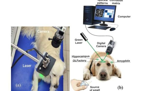
A pioneering study investigating the brain activity of dogs during scent detection has unveiled crucial insights into their remarkable olfactory capabilities. Researchers at Bar-Ilan University have developed an optical sensor capable of remote sensing dogs’ brain activity in three key regions—the olfactory bulb, hippocampus, and amygdala—that play a critical role in how dogs distinguish between different smells. This breakthrough could lead to the development of a compact, non-invasive device capable of interpreting and translating a dog’s olfactory perceptions for human understanding.

In the study published in the Journal of Biophotonics, scientists employed a cutting-edge detection structure system using laser technology and a high-resolution camera to capture brain activity in real-time from four dog breeds.

These dogs were exposed to four distinct scent stimuli—garlic, menthol, alcohol, and marijuana. The data were then analyzed using a machine-learning algorithm revealing that the amygdala plays a significant role in scent differentiation, highlighting the emotional and memory-related aspects of odor processing.

“The findings show that the amygdala is crucial in the way dogs process and react to odors, with specific scents triggering distinct emotional and memory responses, and we are capable of optically detecting their brain activity in this region,” said Prof. Zeev Zalevsky, from the Kofkin Faculty of Engineering at Bar-Ilan University. “This discovery could be the first step toward creating a device that enables us to better understand and interpret the unique way dogs perceive and differentiate smells.”

The study introduces an innovative method of brain activity analysis through laser-based speckle pattern detection, a remote, non-invasive technique that has never been applied to canine brain activity. Unlike traditional methods such as fMRI or EEG, this approach allows researchers to observe brain responses without requiring the dog to be sedated or confined to bulky equipment. This opens up new possibilities for studying dogs in real-world environments, making the technique both affordable and accessible for further research.

Dogs have long been celebrated for their exceptional sense of smell, and this research further illuminates the advanced processes that occur in their brains when detecting odors. With an olfactory system far more developed than humans, dogs can detect a broader range of odors, with specialized receptors in their noses that allow them to process and distinguish even the faintest scents.

This new research offers a glimpse into the intricate workings of the canine brain as it processes different smells, presenting a promising avenue for future applications in areas such as drug detection, medical diagnostics, and search-and-rescue missions.

“Our next step is to develop a portable, Wi-Fi-controlled device equipped with a mini camera and laser system, which could be mounted on a dog’s head and used to monitor its olfactory responses in real time,” said Dr. Yafim Beiderman from Prof. Zalevsky’s Optical Research Lab at Bar-Ilan University.

“This could significantly enhance the way dogs are used in scent detection, from detecting illegal substances to diagnosing diseases in humans, all while deepening our understanding of how they perceive the world around them. More importantly, this real-time sensing could bypass the need to train dogs to utilize their scent abilities.”

The implications of this research could also revolutionize the way dogs are utilized in law enforcement, health care, and beyond. As dogs continue to be invaluable partners in scent detection, this device could provide a means of translating their highly specialized abilities into data that is useful for humans, fostering a stronger connection between the two species.

Current Antivirals, Including Tamiflu, Ineffective Against H5N1 Virus in Cows’ Milk

H5N1 influenza virus continues to circulate in cattle (the virus has been found in dairy cows’ milk and has infected farm workers) causing public health officials to consider preparations for a potential outbreak (or even pandemic) in humans. One aspect of pre-pandemic planning is testing currently approved antivirals against influenza A viruses (like H5N1) circulating in peridomestic species.

In a new study, results suggest that in a preclinical model, two FDA-approved flu antivirals did not successfully treat severe H5N1 infections. Additionally, the researchers found that the route of infection, whether through the eye, the nose, or the mouth, significantly impacts a treatment’s effectiveness.

The findings are published in Nature Microbiology in the paper, “Baloxavir improves disease outcomes in mice after intranasal or ocular infection with Influenza A virus H5N1-contaminated cow’s milk.”

“Our evidence suggests that it is likely going to be hard to treat people severely infected with this bovine H5N1 bird flu strain,” said Richard Webby, PhD, St. Jude Department of Host-Microbe Interactions. “Instead, reducing infection risk by not drinking raw milk and reducing dairy farm workers’ exposures, for example, may be the most effective interventions.”

Though H5N1 infections in people have been rare, there are more than 60 people to date who have become infected from dairy exposures in the current outbreak. Some were infected through exposure to contaminated raw cows’ milk, such as dairy workers who were infected through splashes or aerosolized particles reaching their noses or eyes. Given the risks to human health, the scientists used a mouse model to test how each antiviral drug worked against the virus when it was obtained through three different exposure routes.

“In general, baloxavir [Xofluza] caused a greater reduction in viral levels than oseltamivir [Tamiflu], but neither was always effective,” said Jeremy Jones, PhD, St. Jude Department of Host-Microbe Interactions.

The researchers studied exposure routes that included the eye, mouth, and nose, which are the most common ways to become infected with the virus. The oral route, which mimics drinking raw infected cow’s milk, caused the worst infections that were hardest to treat.

“The virus spread orally far beyond its normal infection of the lungs,” Webby said. “It expanded to the brain and the bloodstream, and the antivirals failed to stop it or improve survival outcomes.”

In contrast, findings showed that baloxavir controlled infections through the eye fairly well. These results are particularly relevant as the ocular route appears to be the common infection pathway for people who work directly with dairy cows. “Baloxavir conveyed 100% survival compared to 25% with oseltamivir,” Jones said. “So, we are seeing enhanced benefits from baloxavir for the ocular infection route.”

Results were mixed for the nasal route. Baloxavir reduced viral levels better than oseltamivir, but the virus still reached the brain. Both antivirals increased survival, with baloxavir and oseltamivir achieving a 75% and 50% survival rate, respectively.

“We showed our existing antivirals’ effectiveness against H5N1 bird flu is route and drug dependent, in some cases doing almost nothing,” Webby said. “Therefore, while we explore different drug combinations and doses, we need to do anything we can to reduce the risk of infection, as that is the best way to protect people from this virus right now.”

Emulsion technique boosts plant growth with bacteria

Researchers have demonstrated a technique for successfully encapsulating bacteria that can then be stored and applied to plants to improve plant growth and protect against pests and pathogens. The technique opens the door to creating a wide range of crop applications that allow farmers to make use of these beneficial bacteria in conjunction with agrochemicals. The paper, “Pickering Emulsion for Enhanced Viability of Plant Growth Promoting Bacteria and Combined Delivery of Agrochemicals and Biologics,” is published in the journal Advanced Functional Materials.

“Many of the beneficial bacteria we know of are fairly fragile, making it difficult to incorporate them into practical, shelf-stable products that can be applied to plant roots or leaves,” says John Cheadle, co-lead author of a paper on the work and a Ph.D. student at North Carolina State University. “The technique we demonstrate here essentially stabilizes these bacteria, making it possible to develop customized probiotics for plants.”

At issue are plant growth-promoting bacteria (PGPBs), which are microbes that benefit plant health and growth, helping plants extract nutrients from the environment and protecting them from pests or pathogens.

“A longstanding challenge for making use of these bacteria has been that if you tried to come up with a single application that combined them with agrochemicals, like pesticides or fertilizers, the bacteria would die,” says Saad Khan, co-corresponding author of the paper and INVISTA Professor of Chemical and Biomolecular Engineering at NC State. “We wanted to develop a solution that would allow bacteria to be used in conjunction with chemicals already in widespread use by growers.”

“By the same token, a healthy plant microbiome allows the plants to make better use of nutrients available in the soil and more resistant to pathogens,” says Tahira Pirzada, co-corresponding author and a research scholar at NC State. “This may allow growers to use less fertilizer and pesticides without hurting crop production.”

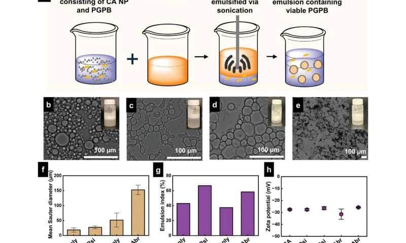
The new technique revolves around a custom-made emulsion, with only a handful of ingredients. One part of the emulsion consists of a saline solution that contains PGPBs. For the proof-of-concept demonstration, the researchers used the bacteria Pseudomonas simiae and Azospirillum brasilense. P. simiae acts as a biopesticide by promoting pathogen resistance; A. brasilense acts as a biofertilizer by fixing nitrogen.

The second part of the emulsion consists of a biodegradable oil and a biodegradable polymer derived from cellulose. The polymer can be loaded with agrochemical active ingredients, which means the emulsion can incorporate these ingredients without relying on environmentally harmful organic solvents, which are typically used in pesticide formulations.

When the two parts of the emulsion are mixed together, the oil is broken into droplets that are distributed throughout the saline solution. The cellulose polymer sticks to the surface of these droplets, preventing the droplets from merging back together.

Essentially, the emulsion is a salad dressing with the oil droplets held in suspension throughout the saline solution. In practical terms, this would allow the PGPBs to be applied simultaneously with agrochemicals using the same emulsion.

To see how well the emulsion worked, the researchers did two tests.

First, the researchers compared the survival of PGPBs in the emulsion to the survival of PGPBs in the saline solution alone. Samples of each were stored at room temperature. After four weeks, the population of P. simiae in the emulsion was 200% higher than the population in saline; the population of A. brasilense in the emulsion was 500% higher.

Engineered E. coli could be used to produce biodegradable plastics

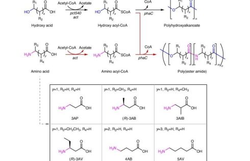
E. coli bacteria could be used to create biodegradable plastics, reports a paper published in Nature Chemical Biology. The engineered bacterial system described in the study may help in the production of plastics with desirable thermal and mechanical properties, using renewable resources, the authors suggest.

Global plastic production is estimated to have created about 400 million metric tons of plastic in 2022, mostly through petroleum-based chemical processes. Meanwhile, the microbial production of polymers has the potential to develop biodegradable alternatives in a more sustainable way.

It is well known that organisms can naturally synthesize polymers, such as DNA, RNA, cellulose and proteins. However, scientists have only recently focused on the use of microorganisms to synthesize polymers that can be used to manufacture plastics.

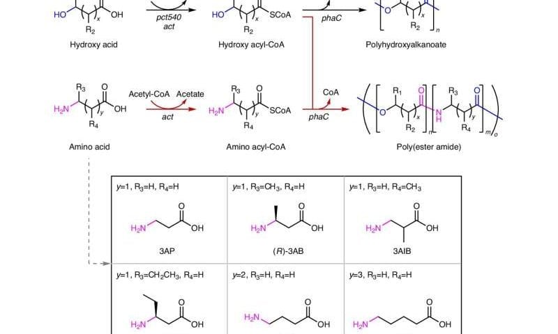
Researcher Sang Yup Lee and colleagues developed a process to produce poly(ester amide) (PEA) using a series of enzymes produced in E. coli; this process involves combining one or more of six amino acids with one or more hydroxy acids to create the polymer plastic. After further tests to optimize the process, Lee and colleagues used glucose as a key ingredient to produce the polymers within E. coli. They also investigated how the amount and structure of the different amino acids used affects the production and properties of the PEAs.

As a proof of concept, the authors produced about 55 grams per liter of a PEA in a large bioreactor, demonstrating that PEA production can be easily scaled up. They also tested the physical, thermal and mechanical properties of this PEA, and suggest that they are comparable to those of high-density polyethylene, one of the most widely used plastics, which indicates that PEAs could serve as a renewable alternative.

This method has several advantages over current chemical methods, such as providing easy access to a wide range of PEAs and enabling the sustainable production of polymers that could be used as plastics, the authors conclude.

The superpower of beans: Resilient, nutritious and ready for climate change

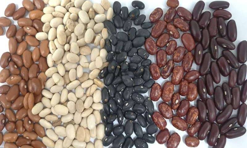
A study conducted by researchers at ESPOL has developed genetically improved bean varieties that are capable of withstanding water scarcity conditions. This discovery is crucial for developing more efficient agricultural strategies. Growing drought-resistant bean varieties will not only ensure stable production but also reduce dependence on excessive irrigation. This promotes more sustainable agriculture.

The work is published in the journal Environmental and Experimental Botany.

Small grain, big impact

Common beans (Phaseolus vulgaris) are a staple food in the diet of many communities worldwide, including Ecuador. Despite their low cost, these small grains are a powerful source of protein, fiber, and essential minerals, making them a key component of food security.

However, bean production is facing increasing challenges due to climate change, particularly water stress. Prolonged droughts affect their growth and yield, thus threatening agricultural sustainability. Identifying proteins involved in drought resistance allows researchers to develop genetic improvement strategies to ensure the viability of crops under adverse conditions.

The hidden language of proteins

What makes some bean varieties more resistant to water scarcity? To answer this question, researchers analyzed nine bean varieties to understand their molecular response to water stress.

Using advanced proteomics techniques, such as two-dimensional electrophoresis (2D-PAGE) and mass spectrometry (MALDI-TOF MS/RP-LC-MS/MS), they identified 111 key proteins involved in drought adaptation. Among them, two proteins stood out for their protective roles: LEA14 and PCC13-62, which help the plant to retain water, stabilize cellular structures, and resist damage caused by lack of moisture.

Notably, the INIAP_473 variety demonstrated exceptional resilience, opening new possibilities for developing crops better adapted to climate change and ensuring food production in a world with less water.

A resilient future for beans and agriculture

The results of this study highlight the importance of biotechnology in agriculture. By gaining a detailed understanding of the proteins involved in water stress resistance, scientists can design more precise breeding programs, optimizing production without compromising food quality.

Moreover, these discoveries benefit not only farmers but society as a whole. A more resilient bean means a more secure food source in times of climate crisis. It also provides an opportunity to reduce the ecological footprint of agricultural production, contributing to water conservation—an increasingly scarce resource.

Thus, the common bean, with its humble appearance, becomes a symbol of resilience and innovation. Through science, we are ensuring that this essential food continues to nourish future generations, even in a world where water is becoming an ever more valuable resource.

How plants could help power the green energy transition

Biological science is changing the way we harness and manage renewable energy, according to a new study by researchers at The Australian National University (ANU).

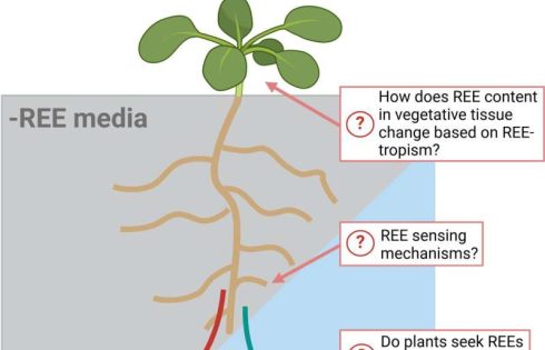
The study, published in Plants, People, Planet, shows how some plant species have evolved to capitalize on the properties of rare earth elements (REEs)—a group of metals essential to the energy transition. Biology has already inspired new metal extraction techniques. For example, “biomining” accounts for approximately 15% of copper mining.

Co-author Professor Caitlin Byrt said we should be looking to biology for inspiration when it comes to efficiently using critical resources, like REEs, to harness and manage clean energy for things like powering our cars and homes.

“Plants are masters of finding efficient strategies for managing and transferring energy. Some plants, for example, exhibit enhanced photosynthesis when they can access REEs,” she said.

“Investigating how plants use these elements in their processes for transferring energy efficiently and precisely could inspire new photon-harvesting devices that mimic photosynthesis, for example.”

REEs are most valuable in their pure forms, which, according to the report’s authors, can be challenging to extract from ores in natural deposits.

“Mechanisms that living organisms use to manage REEs have evolved over millions of years to generate new ways of extracting these valuable resources,” lead author Dr. Samantha McGaughey said.

“Exploration of how REEs interact with biological processes is important for planning the sustainable management of REEs going forward, as we start to rely more and more on these critical resources.

“We need to look at developing innovative approaches to use, reuse and recycle critical resources like REEs and copper to ensure we’re also minimizing negative impacts on the environment. For example, recycling from secondary sources like waste materials can reduce the need for REE extraction from primary sources, using methods that typically lead to disruption of natural ecosystems.”

Professor Byrt added that while there’s exciting potential for using REEs’ resources to manufacture cutting-edge technologies for the future, prioritizing the responsible use of these critical resources is important.

“This means prioritizing the use of critical resources in technologies that improve the quality of life for our communities,” she said.

“Further work in this area of plant science, supported by the Australian Research Council, is expected to help pave the way forward for sustainable use of the resources needed for the clean energy transition.”

The Sustainability Balance in Bioprocessing

The sustainability of bioprocessing depends on many decisions and technologies. As the UK Bioindustry Association (BIA) puts it: “As the world becomes increasingly focused on environmental responsibility, the bioprocessing industry is transforming its approach to reduce its environmental footprint without compromising quality or safety.” In short, sustainability and other factors must be in balance in bioprocessing.

In some cases, scale impacts sustainability. One example comes from Thermo Fisher Scientific. Their DynaDrive single-use bioreactor comes with a 3,000- or 5,000-L bioreactor tank. So, Thermo Fisher compared the sustainability of producing a 5,000-L batch with the DynaDrive using a 5,000-L tank versus a 2,000-L single-use bioreactor. By using the DynaDrive, the process produced nearly 30% less packaging waste.

Thermo Fisher is far from the only company that’s seeking more sustainable solutions for bioprocessors. The BIA site noted above, for example, includes links to sustainability projects at several multinational pharmaceutical companies.

Instead of focusing on just one part of bioprocessing, though, experts must think deeply about various questions. In general, assessing the sustainability of bioprocessing, and most other industries, requires exploring the options, sometimes looking across an entire bioprocess. For instance, two scientists from Sartorius recently noted: “End-to-end continuous bioprocessing can significantly reduce the [cost of goods], increase manufacturing robustness, and reduce the ecological footprint of biopharmaceutical manufacturing of mAbs.”

Plus, even something that sounds less sustainable, like single-use technology, might not be. As Debashis Dutta, PhD, a lecturer in the department of food processing technology at Mirmadan Mohanlal Government Polytechnic in West Bengal, India, and his colleagues pointed out: “As technology progresses, single-use bioreactors are projected to play an increasingly crucial role in developing sustainable and efficient food production processes.” The same could be true for producing biotherapeutics.

Although we live in a world dominated by soundbites and oversimplification, the take-home message is this: Most things are rarely as simple as they seem and only a thorough investigation and data produce actionable information. Plus, the ultimate success of any industry depends on finding balance among competing objectives.

Macrophages Fight COVID-19-Induced Lung Damage with Peroxisomes

Researchers at the University of Virginia (UVA) School of Medicine have uncovered a potential path to treatment for persistent COVID-19 symptoms impacting the lungs. The work, published in Science in an article titled, “Macrophage peroxisomes guide alveolar regeneration and limit SARS-CoV-2 tissue sequelae,” identifies how the viral infection damages macrophages and suggests treatment with a pre-existing FDA-approved drug.

“Our discovery is important because it not only explains why some people continue to have breathing problems long after their initial illness but also points us toward a potential treatment to help them recover,” said senior researcher Jie Sun, PhD, of Carter Center for Immunology Research and the Division of Infectious Diseases and International Health at UVA.

Infection of the respiratory system by diseases like COVID-19 and flu causes damage to the epithelium. During the healing process, regenerative cells are typically kept in check by macrophages, as excessive presence of progenitor cells can lead to pathological tissue remodeling or fibrosis. Macrophages are critical for this process, directing tissue repair through the function of peroxisomes, which reduce inflammation and promote tissue regeneration by digesting lipids and toxins.

“COVID-19 can leave the lungs unable to heal properly by damaging these tiny structures inside our cells,” Sun said.

The team determined that COVID-19 and flu infection damages peroxisomes, thus reducing recovery and prolonging symptoms. In effect, the virus not only causes acute damage but also impairs the body’s ability to heal itself, leading to long-term complications.

“We are collaborating with scientists and physicians at UVA and other institutions to understand the exact function of this understudied organelle in long COVID and other chronic lung diseases such as interstitial lung disease [ILD],” Sun said.

Using a mouse model, the researchers examined changes to macrophage morphology following infection with SARS-COV-2. Mice with severe infection had increased interferon signaling, which both prevented the creation of peroxisomes and accelerated their degradation. This dysfunction resulted in disruption of the macrophage’s ability to function properly, leading to persistent inflammation and lung scarring, key contributors to long COVID symptoms.

In human patients with post-acute sequelae, or long COVID, researchers identified a parallel phenotype of chronic peroxisome impairment as seen in mice. This suggests a similar mechanism in mouse models and humans.

The team further identified a treatment for these chronic symptoms by restoring peroxisome function. Treatment of mouse models with sodium 4-phenylbutyrate (4-PBA) “restored peroxisome function in macrophages, mitigated lung inflammation and fibrosis, and enhanced alveolar regeneration after viral infection” the authors wrote.

The discovery of peroxisomes’ role in controlling inflammation and aiding in lung tissue repair suggests that targeting treatment to these organelles could be a valuable strategy for mitigating chronic lung damage across a range of respiratory illnesses.

“Ultimately, we want to develop peroxisome-targeting therapies to give patients the chance to breathe more easily again and get back to their normal lives,” pointed out Sun.

Additional research is needed before these therapies are used in human patients, but the parallels between human and mouse inflammation and macrophage response are promising. Further, 4-PBA is already an FDA-approved drug for humans, currently used to treat increased levels of ammonia in the blood. Utilizing drugs for new purposes may offer relief to those suffering from long COVID and other persistent post-infection respiratory symptoms sooner than typically expected.

DNA Nanostructures That Form Without Extreme Heat Could Boost Drug Delivery, Diagnostics

Scientists from the RNA Institute at the University of Albany (UAlbany) have developed new methods for designing and assembling DNA nanostructures that enhance their potential for use in various applications from medicine to materials science to data storage. Specifically, they can assemble these structures without the extreme heat and controlled cooling that is typically required.

They also showed that they could assemble these structures in several unconventional buffers including substances like nickel. Full details of the work are published in a new Science Advances paper titled, “Counter ions influence the isothermal self-assembly of DNA nanostructures.”

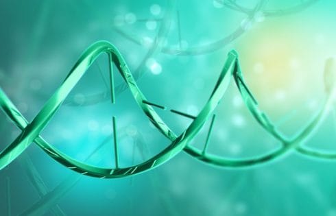
While DNA is most commonly recognized for its role in storing genetic information, it also works well for constructing nanoscale objects. Scientists can create precise structures as small as a few nanometers by programming DNA base pairs and engineering them into shapes with intricate architectures. These structures can be used to accurately place things like biomolecules, cells, and nanoparticles in the context of drug delivery and other use cases.

To create these structures, scientists usually have to heat and cool DNA strands in special buffer solutions that typically contain magnesium ions. This need for precise temperature control limits its use for practical applications in biomedicine for example. Also, DNA nanostructures assembled in magnesium can be unstable in biological environments.

The UAlbany team’s approach offers a way to assemble DNA nanostructures at moderate temperatures using metal ions other than magnesium. “We typically assemble DNA nanostructures by mixing the component DNA strands in a buffer solution, heating the solution to high temperatures, then cooling it down to lower temperatures,” explained Arun Richard Chandrasekaran, PhD, senior author on the study and a senior research scientist at the RNA Institute. With our approach, “DNA nanostructures can be assembled isothermally, that is, at constant moderate temperatures around 68°F or 98.6°F.”

Since their method does not require thermal cyclers or other heating equipment, “it simplifies the process of nanostructure synthesis and opens up the possibility of assembling these structures at constant temperatures,” he said.

Chandrasekaran and his colleagues have previously demonstrated the feasibility of using ions other than magnesium for nanostructure assembly including calcium, barium, sodium, potassium, and lithium at high temperatures. In the current study, they demonstrated that nickel and strontium could also be used with the important distinction that these ions work at moderate temperatures.

Being able to assemble DNA nanostructures at moderate temperatures will make it easier to construct DNA nanodevices for drug delivery and diagnostics using temperature-sensitive proteins like enzymes and antibodies. “Importantly, this work brings us closer to imagining how these nanostructures could actually be made and used in the human body for things like targeted drug delivery or precision diagnostics,” Chandrasekaran said. “While we still have a long way to go before this is possible, demonstrating DNA nanostructure assembly at body temperature is a promising step.”

Extrachromosomal DNA Carrying MYC Drives Pancreatic Cancer Aggression

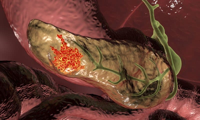
Pancreatic cancer is one of the deadliest cancers worldwide, with a five-year survival rate of 13%. The poor prognosis is due in part both to late detection and the cancer’s capacity to adapt and resist therapy. Laboratory studies by researchers at the University of Verona, the University of Glasgow, and the Botton-Champalimaud Pancreatic Cancer Centre, have now implicated extrachromosomal DNA (ecDNA) carrying the MYC oncogene as a hidden driver of this adaptability.

“Pancreatic cancer is often called a silent killer because it’s hard to detect until it’s too late,” said Peter Bailey, PhD, director of translational research at the Botton-Champalimaud Pancreatic Cancer Centre. “We know that part of its lethality arises from the ability of tumor cells to ‘shape shift’ under stress. Our study shows that ecDNA forms a big part of that story.” Bailey is co-corresponding author of the team’s published paper in Nature, titled “MYC ecDNA promotes intratumor heterogeneity and plasticity in PDAC.” In their paper the researchers’ stated, “Collectively, our work establishes MYC ecDNAs as a key driver of genomic plasticity in PDAC, where they promote rapid and flexible adaptation by amplifying oncogenes, creating heterogeneity, and enabling reversible phenotypic changes.”

Intratumor heterogeneity and phenotypic plasticity drive tumor progression and therapy resistance, the team wrote. However, “… the genetic mechanisms underlying phenotypic heterogeneity are still poorly understood.” For their newly reported study, the investigators sequenced a large panel of patient-derived organoids (PDOs), finding that some pancreatic cancer cells gain a major survival edge by carrying copies of critical cancer genes—such as MYC—on circular pieces of DNA that exist outside of the chromosomes that house most of our genetic material.

These ecDNA genetic rings are free in the cell nucleus, enabling tumor cells to swiftly ramp up gene expression, change their shape, and survive in otherwise hostile environments. The researchers discovered ecDNA to be surprisingly common in pancreatic tumors, particularly for oncogenes like MYC, which drives cancer growth and metabolism. “… we provide a detailed analysis of ecDNAs in PDAC,” they noted. “We have demonstrated that ecDNAs are a major source of high-level amplifications in key PDAC oncogenes and a major contributor to MYC heterogeneity in PDAC.”

Elena Fiorini, PhD, co-first author and senior postdoc, explained, “We saw far more variability in MYC copy number when MYC was on ecDNA. Some cells carried dozens—or even hundreds—of extra MYC copies, giving them a large growth advantage under certain conditions.” The authors further noted, “PDOs and tissues harboring MYC on ecDNA displayed significant heterogeneity of MYC copy number and expression, compared with tumors having MYC on chromosomal DNA.”

Such flexibility underscores the profound intratumor heterogeneity characteristic of pancreatic cancer, where myriad sub-populations coexist and respond differently to treatment. Targeting one subset often fails against another, fueling resistance. “… Overall, we found a heterogeneous landscape of genomic amplifications in PDOs and that ecDNA tumors display features of more biologically aggressive disease,” the team pointed out.

Added Daniel Schreyer, co-first author and a former University of Glasgow PhD student, “It’s effectively a ‘bet-hedging’ strategy. You get pockets of cells that carry very high MYC levels, which is beneficial under certain conditions, and others with fewer copies, which might do better in another environment—all within the same tumor.”

A key advantage of this study is that the organoids—mini-3D replicas of pancreatic tumors grown in the lab—were derived directly from patients with early-stage disease. These organoids preserve much of the genetic make-up of the original tumor, making them excellent testbeds for studying cancer. Unlike methods that artificially introduce ecDNA, these lab models reflect genuine ecDNA variants found in real tumors.

“This approach offers real-world insight into how dynamic and disordered a tumor can be,” said Fiorini. “We see firsthand that even when two patients both have MYC on ecDNA, the structure of that circular DNA can differ substantially—leading to big variations in MYC expression.”

To see how ecDNA drives adaptation, the researchers grew patient-derived organoids and removed vital growth signals—such as WNT factors—and then observed how these organoids responded to the stress. “We found that organoids bearing MYC on extrachromosomal DNA could shift their dependency on WNT,” explained Antonia Malinova, PhD, co-first author and a former PhD student at the University of Verona. “Essentially, cells with high levels of ecDNA became more self-sufficient, no longer needing those external signals to survive.”

The authors stated, “Our analysis revealed that MYC amplification on ecDNA provides a deterministic mechanism for rapid environmental adaptation. A WNT-depleted culture environment drove the rapid selection of cells carrying from dozens to hundreds of ecDNA molecules that could proliferate independently of stromal signals.”

The study also revealed a clear link between high MYC levels and changes in tumor cell shape and behavior. When MYC ecDNA levels soared, cells morphed into more aggressive, solid structures—losing their more organized, gland-like architecture. “Here, we show that extrachromosomal DNA (ecDNA) is a major source of high-level focal amplification in key oncogenes and a major contributor of MYC heterogeneity in pancreatic ductal adenocarcinoma (PDAC),” the investigators stated.

“What’s remarkable,” said co-corresponding author Vincenzo Corbo, PhD, from the University of Verona, “is how rapidly these ecDNA-based copies can appear or disappear depending on the environment. If the cancer is under pressure—say, lacking key growth factors—cells with ecDNA can crank up MYC expression to survive. But if that pressure lifts, they can lose some of these extra DNA circles to avoid the downsides of carrying too many copies.”

Indeed, expressing MYC at high levels can trigger DNA damage, forcing cancer cells to carefully balance the costs and benefits of ecDNA retention. “That was unexpected,” said Corbo. “It challenges the assumption that more MYC is always better for a cancer cell—there’s a real fitness cost to maintaining such high levels.”

Although extrachromosomal DNA only appears in about 15% of patient samples in the reported study, that subset might be particularly aggressive or prone to therapy resistance. As a result, detecting or disrupting ecDNA could open new therapeutic windows. “We might imagine a strategy that exploits the vulnerabilities introduced by ecDNA,” noted Corbo. “Perhaps pushing cancer cells to dial MYC up to a point where they can’t handle the DNA damage, or blocking the molecular circuits that maintain these DNA rings so cells lose them altogether.”

However, the authors caution that such ideas remain early-stage. “ecDNA is a double-edged sword—helpful for quick adaptation but costly to maintain,” Corbo pointed out. “The challenge is to tip that balance in favor of the patient.”

The new work broadens our understanding of genomic plasticity—challenging the notion that the genome is always “fixed.” “We knew the tumor’s surroundings could drive changes, but not that WNT signaling could so directly rewrite DNA,” Bailey commented. “We assumed we’d see mostly epigenetic shifts, so seeing this level of genomic re-engineering was definitely a surprise.”

And with pancreatic cancer cases projected to rise in the coming years, insights into ecDNA’s role could guide future strategies to intercept or exploit this genetic feature—potentially making tumors more vulnerable to treatment.

error: Content is protected !!