Super-resolution microscopy provides real-time picture of bacteria degrading biomass with enzyme complexes

To the untrained eye, they look like blobs blotching the otherwise smooth surface of rod-like bacteria. But if you ask a microbiologist about “cellulosomes,” they will likely tell you that those blobs are actually sophisticated cellulolytic machines.

“Only about 10 to 20 microbes have been so far identified with cellulosomes and characterized, so it is a really rare biomass deconstruction mechanism in nature,” said Yannick Bomble, a National Laboratory of the Rockies (NLR) group manager and senior bioenergy researcher who has studied these enzyme structures for years. “C. thermocellum is one of them, and it is the best biomass degrader that has been identified so far. There’s nothing else that comes close.”

Cellulosomes are big protein complexes packed with dozens of unique enzymes. For years, researchers have known that they are core to C. thermocellum’s exceptional ability to degrade raw biomass. But researchers have lacked a functional picture of how bacteria produce and use cellulosomes—a missing clue to optimize C. thermocellum for turning biomass into chemicals and fuels.

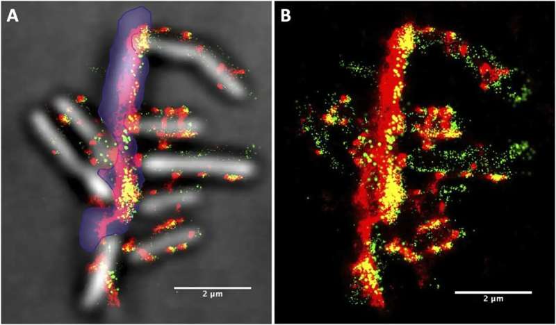
Now, in a paper published in Life Science Alliance, NLR researchers and collaborators used super-resolution imaging and machine-learning-based analysis to quantify the location and movement of cellulosomes on C. thermocellum during biomass deconstruction. The team was surprised not only by the abundance of cellulosomes on each bacterium but also by evidence suggesting that the cells dynamically redistribute them to increase their interaction with biomass.

The team’s machine-learning-guided microscopy techniques represent an advantageous approach for further research—unlocking more insights for someday harnessing C. thermocellum in next-generation consolidated bioprocessing systems.

Large-scale image analysis reveals how C. thermocellum reorganizes cellulosomes
Conventional microscopy is labor intensive, and, as senior NLR researcher John Yarbrough is well aware, it only provides a limited snapshot into the life cycle of bacteria.

“Many microscopy studies capture only a handful of data points,” explained Yarbrough, who is the first author on the paper. “The key question is whether those data points truly represent the full sample and the broader biological system.”

Aiming for a more comprehensive understanding of C. thermocellum, the team, which also included NLR researchers Neal Hengge, Qi Xu, Samantha Ziegler, Daehwan Chung, and Shu Huang, employed an advanced microscopy and analysis technique.

Using a super-resolution microscopy setup—now a well-established capability at NLR that goes beyond traditional optical microscopy to resolve objects barely 30 nanometers apart—they took more than 15,000 images of bacterial samples obtained at different stages of growth. When captured in close succession every 30 milliseconds, each image recorded different signals emitting from the bacteria and cellulosomes, which the team had tagged with fluorescent probes.

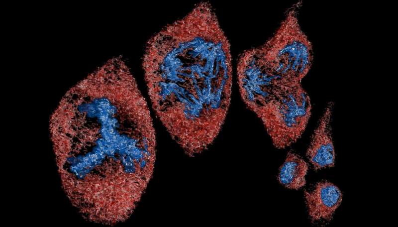
Then, the team used an unsupervised machine-learning clustering algorithm, which acts like a digital organizer to autonomously discover hidden patterns, to analyze the location of each signal. The result was a handful of pictures superimposing hundreds of these signals in a single frame—each showing the location and evolution of cellulosomes during different stages of bacterial growth.

“We noticed that there are significantly more cellulosomes on the plant biomass (i.e., lignocellulose) than on the actual bacteria toward the end of the bacteria’s life,” Yarbrough said. “Where the bacteria were binding with the biomass substrate, we measured in some locations over 1,000 cellulosomes tightly bound in a tiny space.”

In other words, the team resolved the spatial distribution of cellulosome clusters and found that they became enriched at the cell surfaces touching the biomass. As biomass deconstruction progressed, the team noticed that the cellulosomes moved—not remaining fixed on the cell surface. They appeared to disengage, and the bacteria became depleted in cellulosomes in later stages of the cellular life cycle.

More than a pretty picture: A statistical look for quantifying improvements

“What’s exciting to me is that, for once, this imaging is quantitative,” Bomble said of the results. “We can build histograms and conduct statistical analysis to actually measure changes in different stages of bacterial growth, see the size of cellulosome clusters, and measure the number of cellulosomes per unit area.”

For a bacterium like C. thermocellum, such quantitative data provide a basis for someday further engineering them. In the context of industry, the study provides ambition for “consolidated bioprocessing,” where bio-based chemicals and fuels are made in a single pot—rather than two or three using conventional methods.

“The traditional way of achieving biomass deconstruction and its upgrading is pretreatment, enzymatic hydrolysis, and conversion of those sugars into products,” Bomble explained. “What’s appealing about consolidated bioprocessing is that there is no biomass pretreatment or added enzymes, which are significant expenses in cellulosic biofuels, so consolidated bioprocessing could be a lot cheaper.”

Through the Center for Bioenergy Innovation, NLR and other national laboratories are connecting the dots between fundamental microbiology traits and improvements needed for consolidated bioprocessing to work. Thanks to the tools developed in this study, the team was able to connect a few more dots for cellulosomes, which Yarbrough said will also facilitate research on other promising bacteria and microbial consortia.

“This technique allowed us to quantify information to compare results as we genetically modify the cellulosomes,” Yarbrough added. “Do we see change during the growth phases? The beauty of this technique is that we can now go back and compare previous data and show in future publications whether the changes we are making to the cellulosomes lead to improvements in performance.”

NLR’s previous research on cellulosomes has already resulted in intellectual property for biotechnology companies, most recently in another advanced method called cell-free biomanufacturing. Bomble and his team have leveraged the components of the cellulosome to array metabolic enzymes for advanced enzyme immobilization—an example of U.S. Department of Energy Office of Science support resulting in breakthroughs for more applied research.

After all, as Bomble knows, cellulosomes are no mere blobs. Cellulosomes are sophisticated, and the microbes that use them hide insights that could improve how chemicals and fuels are made in the United States.

“This microbe is full of surprises and inspiration,” Bomble said. “It is where ideas come from. This is how fundamental science is now making its way into the more applied sciences.”

Water-based nanocrystal provides a sticky solution to a pesky agricultural problem

A water-based formulation developed at the University of Waterloo using nanotechnology is both greener and more effective than conventional methods for delivering agricultural pesticides.

The new solution dramatically improves how pesticides stick to plant leaves—even in wind and rain—minimizing splash and runoff that contribute to costly waste and environmental contamination.

The study, “Self-assembly of cellulose nanocrystals for splash suppression and enhanced pesticide delivery on hydrophobic surfaces,” is published in the Journal of Colloid and Interface Science.

In early field trials in cabbage plots with an industrial partner in Singapore, the formulation outperformed conventional delivery systems, which use chemicals and solvents to help droplets stick to leaves, by providing better pest control with less pesticide.

“With our new formulation, the pesticide is dispersed in water,” said Dr. Michael Tam, a chemical engineering professor at Waterloo. “We are spraying water, not solvent, making this approach well aligned with sustainable agriculture goals.”

How to effectively deliver pesticides to crops is a major challenge in agriculture. Liquids are now typically applied via nozzles and misting sprays and from planes.

Those methods often fail to deposit pesticides precisely where they are needed. Droplets can bounce off leaf surfaces, drift away in the air, or wash into soil and waterways.

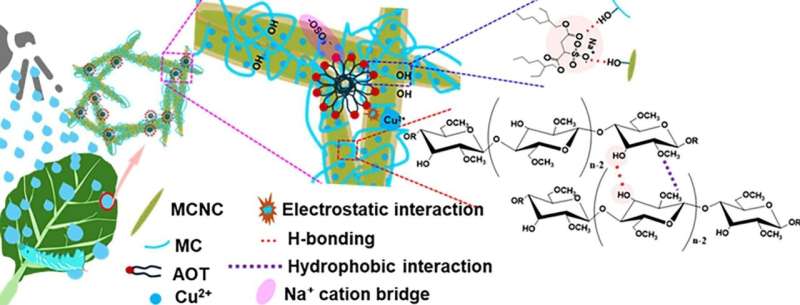
To help solve those issues, Waterloo researchers carefully altered the surface of particles known as cellulose nanocrystals (CNCs) to create a nanostructured formulation that stabilizes pesticide droplets without chemicals or solvents. The formulation uses carbon-neutral CNCs produced from water, pesticides, and inorganic and metallic nanoparticles.

“This unique nanostructure significantly enhances droplet strength, suppressing droplet splash during the impact process,” said Tam, noting high-speed imaging confirmed their effectiveness.

Instead of splashing, fragmenting, or rebounding off leaves, droplets remain intact upon impact, flattening into a pancake-shaped film that adheres strongly. The stabilization method works even when surface tension is reduced by rain and wind, a key distinction from existing technologies.

Researchers are now seeking industrial partners to scale and commercialize their innovation.

When a PDUFA date approaching appears on the pharmaceutical calendar, it represents far more than just another regulatory milestone. The Prescription Drug User Fee Act (PDUFA) date marks the FDA’s commitment to complete its review of a new drug application, creating a pivotal moment that can transform patient treatment options and generate substantial market movements for investors tracking biotech stocks.

The significance of a PDUFA date approaching extends beyond the immediate approval decision. For patients suffering from conditions with limited treatment options, these dates represent hope for breakthrough therapies that could dramatically improve quality of life or even provide life-saving interventions. Pharmaceutical companies invest billions of dollars and decades of research into bringing new medications to market, and the PDUFA date serves as the culmination of extensive clinical trials, regulatory submissions, and scientific review processes.

Understanding the mechanics behind PDUFA dates reveals why they carry such weight in both medical and financial circles. When the FDA accepts a New Drug Application (NDA) or Biologics License Application (BLA), they assign a PDUFA date that typically falls six to ten months later, depending on whether the application receives standard or priority review status. Priority review, reserved for drugs addressing unmet medical needs or offering significant improvements over existing treatments, accelerates the timeline and often signals higher approval probability.

For investors, a PDUFA date approaching creates a defined catalyst event that can trigger significant price volatility. Successful FDA approval often results in substantial stock price increases, particularly for smaller biotech companies where a single drug approval can validate their entire pipeline and business model. Conversely, complete response letters or outright rejections can devastate stock valuations, making PDUFA dates high-stakes events for portfolio managers and individual investors alike.

The patient impact cannot be overstated when considering why these regulatory dates matter so profoundly. Rare disease communities often organize around specific PDUFA dates, advocating for approval of treatments that represent their only hope for effective therapy. Patient advocacy groups frequently submit testimonials and data to the FDA during the review period, highlighting the real-world impact of potential approvals on daily life, symptom management, and long-term prognosis.

Market dynamics surrounding a PDUFA date approaching create unique opportunities for different types of investors. Value-oriented investors might seek undervalued companies with high approval probability, while momentum traders focus on the immediate price action surrounding the announcement. Institutional investors often hedge their positions or adjust allocations based on PDUFA date outcomes, recognizing that these binary events can create or destroy billions in market capitalization within hours of FDA decisions.

The broader healthcare ecosystem also responds to approaching PDUFA dates. Insurance companies begin preliminary coverage assessments, healthcare providers prepare for potential treatment protocol updates, and competing pharmaceutical companies adjust their development strategies based on anticipated approvals. This ripple effect demonstrates how individual drug approvals can reshape entire therapeutic areas and influence treatment standards across the medical community.

Recent regulatory trends suggest the FDA is maintaining rigorous safety and efficacy standards while recognizing the urgent need for innovative treatments. This balanced approach means that a PDUFA date approaching represents a thoroughly vetted evaluation process, giving both patients and investors confidence that approved medications have undergone comprehensive scientific review. The agency’s commitment to meeting PDUFA deadlines also provides predictability in an otherwise uncertain drug development landscape.

As PDUFA dates continue to serve as critical inflection points in pharmaceutical development, their influence on patient outcomes and investment returns remains profound. These regulatory milestones represent the intersection of scientific innovation, patient advocacy, and market dynamics, creating moments where regulatory decisions can literally change lives while simultaneously reshaping investment portfolios. For anyone following the biotechnology sector, understanding the implications of approaching PDUFA dates provides essential insight into both the human impact of medical innovation and the financial opportunities that emerge from successful drug development programs.

Breaking Down How NDA Submission Creates Unprecedented Value for Patients and Investors

The pharmaceutical industry operates on a foundation of regulatory milestones, but few events carry the transformative potential of a successful NDA submission. This critical juncture in drug development represents far more than bureaucratic paperwork—it signals the culmination of years of research, clinical trials, and strategic planning that can fundamentally alter treatment landscapes and investment portfolios alike.

When biotechnology companies reach the point of NDA submission, they’ve crossed a threshold that separates promising research from potential market reality. The New Drug Application process represents the FDA’s most comprehensive review mechanism, requiring companies to present exhaustive data on safety, efficacy, manufacturing, and proposed labeling. This rigorous evaluation transforms experimental therapies into potential life-changing treatments for patients who may have exhausted existing options.

For patients battling serious conditions, an NDA submission often represents renewed hope. The process signals that a therapy has successfully navigated Phase III clinical trials, demonstrating not only safety but also meaningful therapeutic benefit compared to existing standards of care. Oncology patients, for instance, closely monitor NDA submission announcements from companies developing breakthrough cancer therapies, understanding that regulatory filing could mean access to treatments that extend survival or improve quality of life within months rather than years.

The investment implications of NDA submission are equally profound. Biotechnology stocks frequently experience significant volatility around regulatory milestones, with successful submissions often triggering substantial price appreciation. Institutional investors recognize that companies advancing to NDA submission have de-risked their development programs considerably, moving from speculative ventures to entities with quantifiable regulatory pathways toward commercialization.

Market dynamics surrounding NDA submission have evolved significantly as the FDA has implemented expedited review pathways for breakthrough therapies and unmet medical needs. Priority Review designations can compress standard review timelines from twelve months to eight months, accelerating both patient access and investor returns. Companies that successfully navigate NDA submission while securing Priority Review status often see their market valuations reflect this accelerated timeline to potential approval.

The strategic timing of NDA submission also influences competitive positioning within therapeutic areas. First-mover advantage in emerging treatment categories can establish market dominance that persists even after competitor approvals. Companies that achieve NDA submission ahead of rivals often secure favorable positioning with key opinion leaders, payers, and healthcare systems, creating sustainable competitive moats that benefit both patients through improved access and investors through market share protection.

Beyond individual company impact, NDA submission activity serves as a barometer for innovation within specific therapeutic areas. Increased submission volumes in areas like neurodegeneration or rare diseases signal growing research momentum that can attract additional investment capital and pharmaceutical partnership interest. This ecosystem effect amplifies the benefits of successful NDA submission beyond single companies to entire therapeutic sectors.

The financial modeling around NDA submission has become increasingly sophisticated, with analysts developing probability-adjusted revenue projections based on indication size, competitive landscape, and regulatory precedent. Companies with multiple programs approaching NDA submission can command premium valuations due to diversified regulatory risk and expanded commercial opportunities. This portfolio approach to drug development reduces binary outcomes that historically plagued single-asset biotechnology investments.

Patient advocacy organizations have also recognized the significance of NDA submission milestones, often coordinating with regulatory authorities to ensure patient perspectives are incorporated into review processes. This collaboration between stakeholders—patients, companies, investors, and regulators—creates a more holistic approach to drug development that prioritizes both therapeutic innovation and sustainable business models.

The convergence of patient need and investor opportunity through NDA submission represents one of healthcare’s most compelling value creation mechanisms. As regulatory pathways continue evolving to accelerate breakthrough therapy access while maintaining rigorous safety standards, successful NDA submission will likely become an even more powerful catalyst for transforming both patient outcomes and investment returns across the pharmaceutical ecosystem.

De‑extinction company says it’s made an artificial egg—if true, it could help save living species

Today’s announcement by Texas-based de-extinction company Colossal Biosciences about a successful hatching of chicks from an artificial egg would represent a major innovation, if the claims can be verified.

The company says its artificial egg supports the full development of bird embryos outside a biological eggshell, without the requirement for supplemental oxygen. The work is part of its plan to “de-extinct” birds, including the giant moa and dodo.

Colossal’s artificial egg could be groundbreaking science and deliver a useful tool for conservation. But its announcement and slick video include no data or peer-reviewed scientific publications, making it difficult to independently assess the claim.

Artificial egg technology, which involves transferring and growing a developing chick embryo outside a natural eggshell, has been around since the 1980s. Live birds have been hatched from these systems before and grown to adulthood.

The technology is currently used for research purposes such as studying how embryos develop, how tumors grow, and to create genetically modified chickens. It also has applications for drug and vaccine development.

But several stumbling blocks to the widespread use of artificial eggs persist. To improve hatching efficiency, pure oxygen needs to be directly supplied to the developing embryo. This is a double-edged sword because it can also affect chick viability.

Colossal claims to have solved this problem by replacing the hard eggshell and membrane separating the yolk from the shell. Its version is based on the key innovations of an open, latticed half-shell and a transparent, silicone-based membrane that allows oxygen to freely diffuse from the air into the developing embryo.

The company’s plan is to transfer a fertilized embryo and yolk from a real egg to their artificial egg, which would then be housed in incubators. Embryo development would be observed directly through the transparent membrane, as in other artificial systems.

A gene-edited emu

Colossal is planning to genetically modify an emu genome to look more like that of a moa (as they did with gray wolves and dire wolves), create an embryo inside an emu egg, and then bring it to term using this new artificial egg.

The technology could also be used in Colossal’s attempts to genetically engineer a Nicobar pigeon to look more like a dodo.

Key to Colossal’s goal is that its artificial egg could be scaled in size.

However, this still requires a fertilized embryo and yolk. Given the large size differences between chicken eggs and emu (up to 12 times bigger) and giant moa (up to 80 times bigger), there is not enough yolk and egg white in any living birds’ eggs to support the development of a giant moa chick.

An egg yolk is a single cell. It will not be as simple as injecting extra yolk into this fragile cell to make it giant.

Bird embryo development is a complex process, unique to each species. A lot happens in an egg and only time will tell whether this new technology reflects natural processes and produces healthy individuals.

But as our work on other extinct species shows, there is also widespread Māori and public opposition in New Zealand to the company’s plans to “de-extinct” the moa for an ecotourism venture.

A potential conservation tool

The company claims its artificial egg technology “has broad applications for the conservation of threatened species.”

Artificial egg technology requires considerable amounts of funding, which Colossal has mobilized from private sources. This is funding that would not have otherwise been available for conservation.

One area where it could make a significant difference is the captive breeding of critically endangered species (such as kākāpō, kakī black stilt and pukunui southern dotteral) for reintroduction into the wild. This is especially true for long-lived and slow-breeding species which tend to produce fewer eggs.

For example, eggs damaged by inexperienced new parents, misadventure or adverse weather events could be rescued into artificial eggs to help developing chicks to survive.

When combined with genome engineering techniques, the use of artificial eggs could help to reintroduce lost genetic diversity or make birds resistant to diseases. The technology may also be able to reverse the impacts of inbreeding on low hatching success in some species.

However, for critically endangered birds with few natural eggs, the development of transgenic birds would be necessary to produce enough chicks.

For example, chickens could provide sperm and egg cells containing genetically modified DNA from a different species. After mating, the fertilized embryo and yolk could be transferred to the artificial egg.

Ethical questions remain about whether such steps should be taken, even if technologically possible.

The use of artificial egg technology in conservation, especially in combination with genome engineering and transgenic birds, would require transparency and increased levels of engagement with Indigenous communities as the kaitiaki (guardians) of endangered species.

It is also vital this technology (and conservation in general) is not privatized. If Colossal’s artificial egg technology is to make a meaningful difference to saving species from extinction, it must be available to conservation organizations in the public sector.

If the technology lives up to the hype, it won’t be a silver bullet or panacea to stopping species declines, but it might just help. In the short term at least, saving species from extinction will still come down to predator control and habitat restoration.

‘The Silence of the Lambs’ introduced the world to forensic entomology—but how much has changed since?

In the early 1990s, crime-loving television audiences could choose mainly between cozy, fictional detective series such as Columbo and Murder, She Wrote. The US docuseries Unsolved Mysteries brought a few real cold-case investigations to light, but coverage of forensic science on screen was still relatively simple.

Then, in May 1991, “The Silence of the Lambs” was released. Based on Thomas Harris’s 1988 novel, this big-budget thriller was darker, more disturbing, and psychologically complex than most crime films of the time.

The protagonist, FBI trainee Clarice Starling (played by Jodie Foster), is a young woman working in a predominantly male environment—who is often underestimated by her colleagues. When she discovers key evidence through a suspenseful process of extraction from a young victim’s mouth, viewers are introduced to a field of criminal investigation they may never have considered before: forensic entomology.

“Some kind of seed pod?”

“No, sir … that’s a bug cocoon.”

Entomology—the scientific study of insects—is one of the oldest branches of the natural sciences. And the application of insects to criminal cases dates back almost as far. In the forensic text The Washing Away of Wrongs (1247), written by Chinese investigator Sung T’zu, flies attracted to traces of blood on a sickle helped identify a murderer.

However, it was not until the late 19th century that forensic entomology was formalized as a scientific discipline—thanks largely to the studies of Jean Pierre Mégnin. Influenced by his experiences on the battlefield, the French vet began investigating which insects were attracted to animal and human remains at different stages of decomposition.

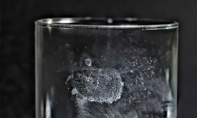
These days, carrion insects are used to tell criminal investigators about the time since a victim’s death, whether their body has been moved, and if any drugs or toxins have contributed to their death.

Human remains are commonly colonized by blowflies and their maggots. But in “The Silence of the Lambs,” Starling was faced with something more unusual: the cocoon of a death’s-head hawkmoth (Acherontia atropos).

The cocoon, which the serial killer inserts into his victims’ throats, is identified by two entomologists who are clearly not forensically trained. Otherwise, they would have thought twice before cutting open the only piece of insect evidence without seeking permission for such a destructive analysis.

The film introduces murderous concepts such as “staging”—the intentional alteration of a crime scene—and a perpetrator’s modus operandi and criminal signature, relating to any distinctive methods they use.

Today, many of us working in forensic entomology and taphonomy (the study of what happens to organisms between death and discovery) are still told our work is “just like ‘The Silence of the Lambs.'” But 35 years after the film’s release, forensic entomology is no longer limited to microscopes, forceps, and entomologists working alone.

Today’s criminal investigations often feature complex interactions between environmental conditions, decomposition processes, and human activity. This makes collaborations between multiple scientific (and non-scientific) disciplines essential.

How the science has progressed

In the two decades preceding the film’s release, the biomedical and life sciences journal PubMed listed 37 publications on the subject of forensic entomology. Since then, there have been more than 1,800.

Methods used for insect identification and age estimation have changed dramatically. Today, molecular and chemical techniques can identify insect species and determine their stage in the lifecycle and geographic origin. These techniques are especially useful in cold cases or poorly preserved crime scenes, where samples may have been damaged or improperly stored.

Insects are also playing an increasingly accurate role in determining the time of death. As well as feeding on decomposing remains, they help spread the bacteria and other microorganisms involved in decomposition. These microbial communities change in predictable ways over time—even in extreme environmental scenarios—offering investigators a further indicator of the postmortem interval.

Chemical profiling of insect cuticular hydrocarbons (insect skin) provides definitive species and age signatures. These can reduce the risk of error associated with identification by people, and the time and costs of DNA sequencing.

Forensic entomology has also expanded into areas such as entomotoxicology, where insects feeding on decomposing remains are analyzed for the presence of drugs, toxins, or other chemical compounds. It is even possible to recover the DNA of the individual on whom an insect has been feeding directly from that insect’s gut contents.

In “The Silence of the Lambs,” investigators assume that “water leaves no trace evidence of any kind.” Yet today, aquatic forensic researchers examine not only insects but crustaceans, microorganisms, and bone proteins associated with decomposing remains in water.

Revisiting the moth cocoon scene

The film’s infamous moth cocoon scene—which saw the extracted evidence collected with forceps, then taken for visual inspection at a museum—would be approached rather differently today.

Firstly, spoons are now recommended over forceps to avoid damaging the sample. Modern forensic practice aims to preserve specimens by taking photographs before any manipulation. Where possible, insects are reared to the adult stage, which is often easier to identify with certainty.

Rather than opening the cocoon, it could be compared as is with museum reference collections or analyzed using technology such as hyperspectral imaging. This would confirm the species and estimate its developmental stage without altering the evidence.

Many high-profile cases, including some wrongful convictions, have demonstrated how forensic entomology can be a key tool in the investigation of current and historical crimes—as well as of natural disasters and war crimes.

However, technological advances are not enough. The reliability of forensic entomology depends on appropriate crime scene protocols, evidence collection, ongoing research and, perhaps most importantly, specialist training and attention to detail. These qualities are certainly embodied by agent Starling.

But there is another major difference since the film came out in 1991. Unlike Starling’s experience, women now represent a major part of the forensic science workforce. They contribute to a discipline that has become far more diverse, collaborative, and scientifically advanced than the one portrayed in “The Silence of the Lambs.”

Hi-res microscopes give biologists petabytes of data. Scientists are creating an AI assistant to make sense of it

In a cramped, windowless room on the University of California, Berkeley, campus, two bespoke microscopes—each a Swiss Army knife for high-resolution imaging—operate around the clock gathering data that will help train a game-changing technology for the field of biology: AI.

The identical microscopes, described this week in the journal Nature Methods, squeeze a dozen types of high-powered microscopes into a single machine, from standard phase contrast to the latest lattice light-sheet technology—easily switchable with the push of a button. Called MOSAIC (Multimodal Optical Scope with Adaptive Imaging Correction), it has already been recreated in more than a dozen labs worldwide thanks to preprints and elaborate assembly instructions disseminated over the past six years.

At UC Berkeley, it is one in a lineup of improved imaging technologies that could forever alter the field of biology, the researchers say. The microscopes can track over seconds, hours, or days the development of live specimens, ranging from molecules and cells to entire embryos, gathering huge amounts of data that will allow biologists to track cells as they move through tissue, the evolution of internal cellular structures, and even the shuttling of proteins and other molecules within the cell.

All this data—measured in petabytes, the equivalent of about 500 billion pages of text—requires the analytic ability of a large “vision” language model (LVLM), like ChatGPT. Building an LVLM or AI that can deal with petabytes of imaging data is now one of the main focuses of a team of microscopists, physicists, biologists, and computer scientists in Berkeley’s Advanced Bioimaging Center, which hopes to create a first-of-its-kind Cell Observatory.

“Life has to be studied in living tissue, holistically, and over fast timescales and for long periods of time,” said Eric Betzig, a Berkeley professor of molecular and cell biology and of physics who won the 2014 Nobel Prize in Chemistry for the development of super-resolution fluorescence microscopy—a version of which is now incorporated into MOSAIC.

“You can’t study something as complex as a cell or organism just by looking at the parts individually—there are something like 40 million protein molecules alone of 20,000 different types. With our microscopes, we can image everything from single molecules to whole organisms at high resolution, following as many players as we can to understand natural physiological interactions in the cell.”

Betzig, a Howard Hughes Medical Institute investigator, refers to the imaging data as five-dimensional, or 5D: three spatial dimensions, plus time and color. The color comes from fluorescent labels that allow scientists to track multiple subcellular structures simultaneously—organelles, membranes, the cytoskeleton, and more—as they migrate, change shape, divide, and interact over time.

“We are the world’s best at collecting data at 5D, and have been for a decade,” he said. “But we don’t know how to interpret the data at scale; we can’t think in petabytes and we don’t see in 5D. That’s why we’re developing a 5D AI—it’s a sherpa to guide us.”

MOSAIC’s development was led by Srigokul “Gokul” Upadhyayula, an assistant professor in residence in molecular and cell biology who had earlier worked with Betzig on other high-resolution techniques, the adaptive optical lattice light-sheet microscope, and the expansion lattice light-sheet microscope—both now part of MOSAIC.

“Biology is entering an era in which the data are too complex and too large to interpret by human inspection alone,” he said. “A biologist may understand the biological question deeply, but still lack the computational tools and infrastructure needed to process, analyze, and quantify what they are seeing. We need to build a mind that can reason natively with 3D movies of living biological systems and let us query those dynamics through language—akin to a ChatGPT for biology.”

One remarkable video they captured shows a zebrafish regrowing its tail fin. Although the movie itself spans only 12 hours, it took months of preparation, processing, and visualization before they could fully understand what it showed. The video revealed tiny events inside living tissue that are normally very hard to see: cells near the wound releasing small communication packets, microscopic fibers beneath the skin shifting as the tissue repaired itself, two repair cells fusing together, and a red blood cell briefly getting trapped as new blood vessels were remodeled.

An AI assistant would not only help assemble these data-intensive movies, but help biologists home in on the specific activities they’re interested in.

“There’s so much information in these large movies, across scales, about how cells are behaving in the organism and the tissue and at the subcellular level, it can be difficult even for a very well-trained biologist to understand or digest,” said Ian Swinburne, a Berkeley assistant professor of molecular and cell biology who works with Upadhyayula’s team to study how cells engulf other cells, such as the macrophages that clean up dead cells in a wound.

“AI can help us interface with the data and ask or answer questions more easily. Like, ‘How many macrophages are crawling into my tissue during an infection?’ or ‘Can I predict when a cell’s going to start leaving its organ?’ That happens in development but also in cancer during metastasis.”

“The impact of MOSAIC will be minimal until we build an AI model to be able to deal with the data that comes out of those systems; we basically have a gold mine, but we have no ability to get the gold out,” Upadhyayula said. “The primary output of our Cell Observatory Initiative will be an AI mind that’s able to be our scientific partner in extracting these observations.”

A ‘Swiss Army Knife’ microscope

MOSAIC relies on several advances, Upadhyayula said, fluorescent molecules that allow biologists to mark specific cellular structures and molecular activities in living cells; fast, gentle light-sheet imaging that captures those dynamics with minimal stress or damage to the cells; high-speed data transfer and computing infrastructure capable of moving and processing massive imaging datasets; and new computational tools, including AI models, to help interpret the resulting 3D movies of living systems.

He and his colleagues in the Advanced Bioimaging Center combine these to create one-off high-resolution microscopes, such as the super-resolution microscope, which won Betzig the Nobel Prize. That innovation involved using a laser to stimulate fluorescent tags, which allowed researchers to image individual molecules in a cell and superimpose them into high-resolution images.

Betzig subsequently developed a faster but gentler technique for hi-res cell imaging, lattice light-sheet microscopy (LLSM), which reduces cellular damage by spreading the laser energy across a thin sheet to more gently illuminate a transparent specimen one thin slice at a time. The fluorescing markers are captured in real time and assembled into a 3D video.

MOSAIC combines these and other high-resolution imaging techniques into a single machine that can quickly transition from one imaging mode to another, repositioning many of the lenses that shape the light. To sharpen images, it uses adaptive optical elements, such as a deformable mirror controlled by 69 tiny motors that make minute adjustments to correct for blurring caused by aberrations in the living tissue itself.

Among the available modes are the latest versions of light-sheet and super-resolution microscopy, as well as multi-photon and label-free imaging. Across the various modes, MOSAIC is able to capture subcellular dynamics in cultured cells and live multicellular organisms, map nanoscale features across millimeter-scale expanded tissues, and image the neural architecture in the brains of live mice.

Movies make a difference

The researchers emphasized the importance of video in understanding biological interactions, and the need to see them with sufficient fidelity.

“The name of the game is to keep the organism, the sample, as physiologically happy as possible,” Upadhyayula said. “Which means using the lowest light dose we can to keep it from deep-frying while getting the information we need. The consequence is that the image gets noisy. When we watch a noisy movie over time, our minds naturally filter out some of the noise and focus on the underlying structures.”

But getting AI to interpret 5D images is significantly harder than conventional image recognition.

“The current vision models are not built to reason over three dimensions, time, and molecular identity or color, and that’s what we want to build,” he said.

Swinburne and Dave Matus, a researcher working with Betzig and Upadhyayula on the Cell Observatory Initiative, are now helping develop new labeling reagents that highlight subsets of the thousands of components for AI to recognize. While thoroughly impressed with MOSAIC’s ability, Swinburne admits that the videos are so good that it’s hard to focus on just one thing.

“There’s so much information in these movies,” he said. “We come in with maybe a hypothesis about the process we think we’re studying and then we get distracted by something we’ve never seen before. Probably every movie has something new that we acquire just because the quality is so high, the spatial and time resolution so much better than what we’re used to.”

The four first authors of the Nature Methods paper are Gaoxiang Liu and Xiongtao Ruan of UC Berkeley and Tian-Ming Fu and Daniel Milkie at the Janelia Research Campus of the Howard Hughes Medical Institute (HHMI) in Virginia. Upadhyayula, Betzig, and Wesley Legant of the University of North Carolina at Chapel Hill are senior authors of the paper. Betzig is a HHMI investigator and Upadhyayula is a Biohub San Francisco investigator.

Agentic AI could help electron microscopes plan, adapt and analyze experiments

Scientific discovery is often portrayed as the result of long hours alone in a lab, but true science is inherently collaborative. The most robust experimental processes are developed through partnerships across multiple areas of research.

The need for specialized, multidisciplinary teams slows experiment design, execution, data analysis, and process updates, delaying technological validation and deployment. But if the increasingly automated tools scientists already use in the lab could contribute to this team process of experimental design, the timeline for these goals could be greatly accelerated.

Introducing thinking electron microscopes

This concept of “lab tool as lab assistant” is the premise of a paper in npj Computational Materials titled “Thinking Microscopes: Agentic AI and the Future of Electron Microscopy,” by Vida Jamali, assistant professor the School of Chemical and Biomolecular Engineering; Amirali Aghazadeh, assistant professor in the School of Electrical and Computer Engineering; and Josh Kacher, associate professor in the School of Materials Science and Engineering.

In the paper, the team introduces the concept of “thinking electron microscopes,” in which agentic AI systems are directly integrated with the instrument. This allows microscopes to move beyond their conventional role as characterization tools and toward functioning as co-scientists for human users.

Drawing on advances in specialized large language models, or LLMs, that demonstrate their ability to collaborate, reason over data, and integrate prior knowledge, the team envisions specialized LLM-based agents assigned to specific roles and areas of knowledge expertise.

By explicitly incorporating domain knowledge into specialized agents and distributing information across multiple agents with focused expertise, the approach enables parallel evaluation of competing hypotheses, clearer separation of roles—such as planning, simulation, and critique—and more transparent and robust reasoning.

How agentic AI supports experiments

Within the experimental pipeline, these agents can analyze materials’ properties, physical data, chemical processes, and other relevant parameters. They could also collaborate with an agent that specializes in experimental design, refining iterative closed-loop experimentation, and real-time scientific discovery.

Although the research focuses on AI collaboration, the team notes that human researchers must retain accountability for the accuracy and integrity of both the experimental process and the results reported.

This oversight begins with advocating for greater open access to research materials in all formats, building community-driven data repositories, and adopting standardization in how experimental parameters and metadata are reported.

Equally important, researchers should be willing to report data from failed experiments as well as successful outcomes. Finally, organizations should work together to standardize secure APIs that enable shared, remote access to infrastructure across distances.

Building the next generation of tools

“We see this as a step toward scientific instruments that do more than acquire data; systems that can reason over experiments, adapt measurements, and participate in the scientific discovery process alongside researchers,” says assistant professor Vida Jamali.

The team is already developing these systems by connecting cloud-based, agentic infrastructures to microscopes at the Institute for Matter and Systems at Georgia Tech.

With the addition of agentic AI, the goal is to accelerate discovery and engineering of new nanoscale materials for energy and quantum applications, as well as advance capabilities in cryo-electron microscopy and structural biology. These tools can optimize data collection, link real-time microscope observations with structural models of proteins, and dynamically adjust and prioritize experiments.

The team sees this work as the first step toward the next generation of “thinking” electron microscopes, as well as an advancement in scientific discovery across domains.

AI-designed miniproteins switch key cell receptors on and off

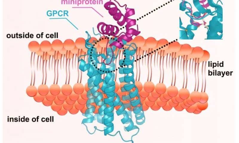
G protein-coupled receptors, or GPCRs, sit in the plasma membrane, the boundary that defines the inside and outside of a living cell. They communicate with nearly every physiological process in our bodies—from the ability to see and smell, to sensing of adrenaline, insulin, nutrients and medicines.

A key challenge has been developing molecules that can toggle GPCRs on and off in different contexts. Doing so would not only provide insight into how these receptors control almost every bodily function, but also inroads to medicines for a host of diseases that lack treatments.

The UW Medicine Institute for Protein Design and Skape Bio led a new study showing for the first time that AI can be used to create computationally designed proteins to activate or block GPCRs.

Their findings are published in Nature.

“Protein design takes our understanding of how proteins fold and reverses it—asking if we can envision, with the aid of AI computing, a new protein that sticks to a target in a purpose-built way,” said senior author David Baker, director of the UW Medicine Institute for Protein Design.

“This paper showcases how we can do this repeatedly for different GPCRs in ways that capitalize on their dynamic motion to either activate or inactivate them. The result is a generalized approach to targeting biologically critical receptors,” added Baker, who is a professor of biochemistry at the University of Washington School of Medicine and a Howard Hughes Medical Institute Investigator.

The signaling switch of GPCRs sits in deep flexible pockets, the shape of which makes them difficult to target. The team developed specialized design strategies to build miniproteins (proteins with fewer than 100 amino acids) that can slip into these hard-to-access sites. This approach enabled the generation of molecules designed to either activate or block signaling.

By targeting specific active or inactive receptor states, the team designed miniproteins that precisely control GPCR signaling in cells, either turning it on or shutting it down. Structural studies showed that several closely matched their design models. In one mouse study, a designed miniprotein performed comparably to a clinically used drug while showing fewer side effects.

“Existing drugs such as antibodies bind to but often fail to activate or block GPCR signaling,” said Edin Muratspahić, an Institute for Protein Design postdoctoral research scholar and a first author of the study. “Seeing computationally designed miniproteins not only bind but actually control GPCR signaling in living cells was a defining moment for me.”

To accelerate the discovery of designed proteins targeting GPCRs, the researchers also invented a new screening system. Traditional screening is difficult for these receptors because many methods require that they be purified, stabilized, or otherwise altered in ways that can change their signaling.

By working directly in living human cells, the new system can test tens of thousands of proteins against GPCRs while keeping the receptors in the cell membrane.

“The methods we are sharing in this new study form the roadmap for achieving all-computational design of protein ligands for any GPCR,” said Christoffer Norn, corresponding author and co-founder of Skape Bio.

“One of the great strengths of the Institute for Protein Design is its capacity to drive its research quickly from the university setting to start-ups that can carry that work forward into real-world impact.”

“At Skape Bio,” added Norn, “we are achieving this impact by maturing the methods and approaches necessary to deliver therapies for patients across a wide range of diseases where GPCRs are known to be effective targets, but where medicines have not previously been available.”

Researchers transform paper sludge into valuable biofuels

Researchers have demonstrated that different types of paper industry sludge, typically treated as a low-value waste, can be transformed into a high-yield renewable biofuel. The study, published in Biofuels, Bioproducts and Biorefining, reveals that paper sludge streams vary significantly in their suitability for producing bioethanol and biogas, offering a pathway to more efficient waste-to-energy strategies.

The research examined three major sludge types generated by the pulp and paper sector: virgin pulp sludge, corrugated cardboard sludge, and tissue and printing paper sludge. Each of these materials was assessed for its composition, enzymatic digestibility, and performance in bioethanol fermentation and anaerobic digestion.

Worldwide, as much as 500 million tons of wet paper sludge are created each year through the paper production process. This waste is fiber-rich, and often ends up in landfill, leading to methane emissions and water loss. As well as minimizing waste, production of biofuels such as biomethane, biohydrogen, and bioethanol from this kind of biomass has potential as a sustainable alternative to fossil fuels.

Virgin pulp sludge was found to deliver the highest bioethanol potential, with corrugated cardboard sludge producing the greatest volume of biogas and methane. The study revealed differences in the biochemical and physical properties of the varieties of paper sludge, which could inform circular bioeconomy initiatives in the paper industry going forward by matching the sludge characteristics with the most efficient bioconversion route.

error: Content is protected !!