Weight-loss, diabetes drugs linked to vision problems in small study

Popular drugs for diabetes and weight loss could have an unexpected side effect.

Glucagon-like peptide 1 (GLP-1) receptor agonists, which are used to treat type 2 diabetes and/or obesity, were linked to vision problems in a small study at the University of British Columbia.

Some common GLP-1 drugs include Ozempic and Wegovy, which contain semaglutide as the active ingredient, and Mounjaro and Zepbound, which contain tirzepatide.

In the study, nine patients who were using a GLP-1 developed “ophthalmic complications,” according to the researchers. The average age of the patients was 57.4 years, according to the study findings.

Seven of the patients had nonarteritic anterior ischemic optic neuropathy (NAION), which causes vision loss in one eye.

One patient developed bilateral papillitis, which involves swollen optic nerves that can cause impaired vision, and another had paracentral acute middle maculopathy, which leads to a blind spot in the retina.

All the patients had a history of type 2 diabetes, hyperlipidemia (high lipids or fats in the blood), hypertension and/or sleep apnea.

The findings were published in JAMA Ophthalmology.

“In one of the cases presented, the patient was taking the drugs for weight loss and did not have a prior history of diabetes (which can also be linked to the condition),” Mahyar Etminan, associate professor of medicine at the University of British Columbia, told Fox News Digital. (Etminan was author of the commentary on the study.)

“In another case, when the drug was stopped and reintroduced, the condition reappeared, strengthening a causal link.”

Ziyad Al-Aly, a clinical epidemiologist at Washington University in St. Louis, was not involved in the study but shared his comments on the findings.

“This is a very small study and it was uncontrolled — meaning it did not include people who were not using GLP-1 drugs,” he told Fox News Digital.

“The story of GLP-1 is still being written — and we are learning something new about these drugs every day.”

“This makes it impossible to know whether the reported eye problems are caused by these drugs.”

Nevertheless, the doctor noted, “the story of GLP-1 is still being written — and we are learning something new about these drugs every day. The findings in this study should be pursued further.”

Etminan also acknowledged the study’s limitations.

“This data was derived from a series of individual cases and was not an epidemiologic study,” he noted. “However, another recent epidemiologic study also confirmed an increase in risk.”

Al-Aly called for large, controlled studies — including people who take the drug and a control group of people who are not using the drug — to evaluate the long-term health effects of these medications, including eye problems.

“In the meantime, for people who may be at risk of vision problems, or who already have vision problems, caution is advised,” he added. “People should discuss with their doctors to determine if GLP-1 is the right medication for them.”

Etminan echoed that cautionary guidance.

“Those taking these drugs for diabetes should probably continue taking them for their cardiovascular benefits, but be aware of the signs of NAION,” he advised.

“Healthy individuals taking them to lose a few pounds for an event might want to more carefully weigh the risks versus the benefits of taking these drugs.”

“Most of the vision side effects appear to resolve when the medication is stopped.”

Dr. Seth Kipnis, medical director of bariatric and robotic surgery at Hackensack Meridian Jersey Shore University Medical Center, noted that there have been “rare and unusual side effects” from this class of medications, but he believes the vision changes seem to be more related to the rapid blood sugar changes caused by the medications than to the medications themselves.

“We have encouraged any patients who are on these types of medications to report any unusual symptoms to their prescribing doctors,” Kipnis, who also was not involved in the research, told Fox News Digital.

“Most of the vision side effects appear to resolve when the medication is stopped.”

Kipnis emphasized that these drugs should only be taken under the care of a healthcare professional and that “good and consistent follow-up for dose adjustments with monitoring for side effects” is critical.

When contacted by Fox News Digital, Novo Nordisk (maker of Ozempic and Wegovy) provided the following statement.

“NAION is a very rare eye disease, and it is not an adverse drug reaction for the marketed formulations of semaglutide (Ozempic®, Rybelsus®and Wegovy®) as per the approved labels. After a thorough evaluation of studies from the University of Southern Denmark and Novo Nordisk’s internal safety assessment, Novo Nordisk is of the opinion that the benefit-risk profile of semaglutide remains unchanged.”

The company also noted that eye conditions are “well-known comorbidities” for people living with diabetes.

“Any decision to start treatment with prescription-only medicines should be made in consultation with a healthcare professional who should do a benefit-risk evaluation for the patient in question, weighing up the benefits of treatment with the potential risks,” Novo Nordisk added.

Enzymes are the engines of life—machine learning could help scientists design new ones

Enzymes are molecular machines that carry out the chemical reactions that sustain all life, an ability that has captured the attention of scientists like me.

Consider muscle movement. Your body releases a molecule called acetylcholine to trigger your muscle cells to contract. If acetylcholine sticks around for too long, it can paralyze your muscles—including your heart muscle cells—and, well, that’s that. This is where the enzyme acetylcholinesterase comes in. This enzyme can break down thousands of acetylcholine molecules per second to ensure muscle contraction is stopped, paralysis avoided and life continued. Without this enzyme, it would take a month for a molecule of acetylcholine to break down on its own—about 10 billion times slower.

You can imagine why enzymes are of particular interest to scientists looking to solve modern problems. What if there were a way to break down plastic, capture carbon dioxide or destroy cancer cells as fast as acetylcholinesterase breaks down acetylcholine? If the world needs to take action quickly, enzymes are a compelling candidate for the job—if only researchers could design them to handle those challenges on demand.

Designing enzymes, unfortunately, is very hard. It’s like working with an atom-sized Lego set, but the instructions were lost and the thing won’t hold together unless it’s assembled perfectly. Newly published research from our team suggests that machine learning can act as the architect on this Lego set, helping scientists build these complex molecular structures accurately.

What’s an enzyme?

Let’s take a closer look at what makes up an enzyme.

Enzymes are proteins—large molecules that do the behind-the-scenes work that keep all living things alive. These proteins are made up of amino acids, a set of building blocks that can be stitched together to form long strings that get knotted up into specific shapes.

The specific structure of a protein is key to its function in the same way that the shapes of everyday objects are. For example, much like a spoon is designed to hold liquid in a way that a knife simply can’t, the enzymes involved in moving your muscles aren’t well suited for photosynthesis in plants.

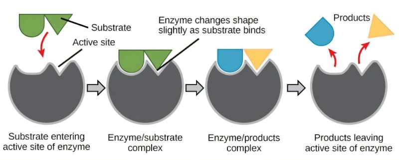
For an enzyme to function, it adopts a shape that perfectly matches the molecule it processes, much like a lock matches a key. The unique grooves in the enzyme—the lock—that interact with the target molecule—the key—are found in a region of the enzyme known as the active site.

The active site of the enzyme precisely orients amino acids to interact with the target molecule when it enters. This makes it easier for the molecule to undergo a chemical reaction to turn into a different one, making the process go faster. After the chemical reaction is done, the new molecule is released and the enzyme is ready to process another.

How do you design an enzyme?

Scientists have spent decades trying to design their own enzymes to make new molecules, materials or therapeutics. But making enzymes that look like and go as fast as those found in nature is incredibly difficult.

Enzymes have complex, irregular shapes that are made up of hundreds of amino acids. Each of these building blocks needs to be placed perfectly or else the enzyme will slow down or completely shut off. The difference between a speed racer and slowpoke enzyme can be a distance of less than the width of a single atom.

Initially, scientists focused on modifying the amino acid sequences of existing enzymes to improve their speed or stability. Early successes with this approach primarily improved the stability of enzymes, enabling them to catalyze chemical reactions at a higher range of temperatures. But this approach was less useful for improving the speed of enzymes. To this day, designing new enzymes by modifying individual amino acids is generally not an effective way to improve natural enzymes.

Researchers found that using a process called directed evolution, in which the amino acid sequence of an enzyme is randomly changed until it can perform a desired function, proved much more fruitful. For example, studies have shown that directed evolution can improve chemical reaction speed, thermostability, and even generate enzymes with properties that aren’t seen in nature. However, this approach is typically labor-intensive: You have to screen many mutants to find one that does what you want. In some cases, if there’s no good enzyme to start from, this method can fail to work at all.

Both of these approaches are limited by their reliance on natural enzymes. That is, restricting your design to the shapes of natural proteins likely limits the kinds of chemistry that enzymes can facilitate. Remember, you can’t eat soup with a knife.

Is it possible to make enzymes from scratch, rather than modify nature’s recipe? Yes, with computers.

Designing enzymes with computers

The first attempts to computationally design enzymes still largely relied on natural enzymes as a starting point, focusing on placing enzyme active sites into natural proteins.

This approach is akin to trying to find a suit at a thrift store: It is unlikely you will find a perfect fit because the geometry of an enzyme’s active site (your body in this analogy) is highly specific, so a random protein with a rigidly fixed structure (a suit with random measurements) is unlikely to perfectly accommodate it. The resulting enzymes from these efforts performed much more slowly than those found in nature, requiring further optimization with directed evolution to reach speeds common among natural enzymes.

Recent advances in deep learning have dramatically changed the landscape of designing enzymes with computers. Enzymes can now be generated in much the same way that AI models such as ChatGPT and DALL-E generate text or images, and you don’t need to use native protein structures to support your active site.

Our team showed that when we prompt an AI model, called RFdiffusion, with the structure and amino acid sequence of an active site, it can generate the rest of the enzyme structure that would perfectly support it. This is equivalent to prompting ChatGPT to write an entire short story based on a prompt that only says to include the line “And sadly, the eggs never showed up.”

We used this AI model specifically to generate enzymes called serine hydrolases, a group of proteins that have potential applications in medicine and plastic recycling. After designing the enzymes, we mixed them with their intended molecular target to see whether they could catalyze its breakdown. Encouragingly, many of the designs we tested were able to break down the molecule, and better than previously designed enzymes for the same reaction.

To see how accurate our computational designs were, we used a method called X-ray crystallography to determine the shapes of these enzymes. We found that many of them were a nearly perfect match to what we digitally designed.

Our findings mark a key advance in enzyme design, highlighting how AI can help scientists start to tackle complex problems. Machine learning tools could help more researchers access enzyme design and tap into the full potential of enzymes to solve modern-day problems.


AI model generates antimicrobial peptide structures for screening against treatment-resistant microbes

A team of microbiologists, chemists and pharmaceutical specialists at Shandong University, Guangzhou Medical University, Second Military Medical University and Qingdao University, all in China, has developed an AI model that generates antimicrobial peptide structures for screening against treatment-resistant microbes.

In their study published in the journal Science Advances, the group developed a compression method to reduce the number of elements needed in training data for an AI system, which helped to reduce diversification issues with current AI models.

Prior research has suggested that drug-resistant microbes are one of the most pressing problems in medical science. Researchers around the world have been looking for new ways to treat people infected with such microbes—one approach involves developing antimicrobial peptides, which work by targeting bacterial membranes.

Unfortunately, developing or finding peptides has proven to be too slow to address the crisis. So researchers have turned to AI-based approaches to aid in finding such peptides. But that approach has encountered problems, as well, the biggest being the lack of a large training base, which leads to peptide discovery that lacks diversity.

In this new study, the researchers in China found a way around this problem by developing a compression technique that reduces the number of elements needed to train their AI system.

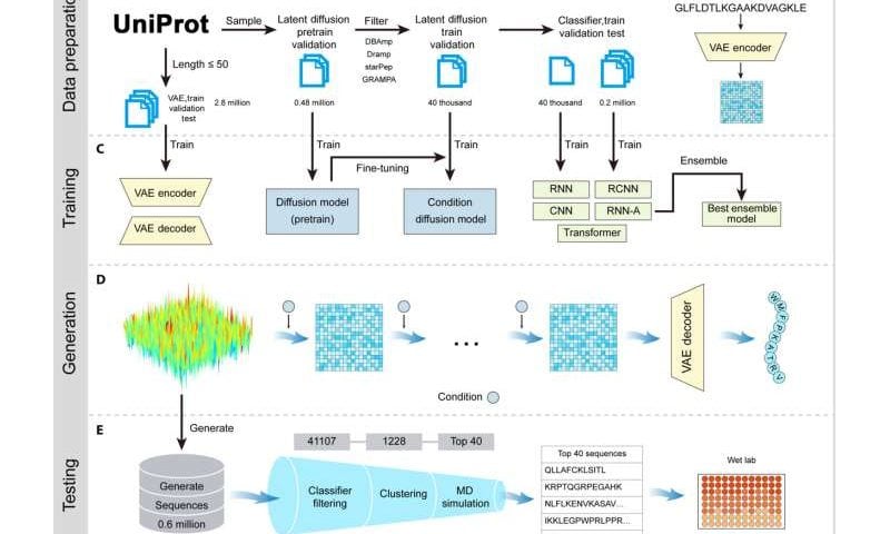
The researchers call their system a two-stage AI pipeline leverage diffusion model. The first stage works by compressing data describing 2.8 million known peptides into a numerical form by amplifying signal noise randomly. The second stage then pulls new peptides from the simplified data, removes the noise, and decompresses the data used to describe its peptide sequence.

In testing their new system, the research team found that it was able to filter peptides listed in a training database down to a reasonable number of those most likely to have antimicrobial properties. In looking at 600,000 of them, the team experimentally tested 40 peptides and found 25 that showed promise in combating bacterial and fungal pathogens.

Electrified Purification Could Eliminate Problematic Buffers

The buffers used in downstream purification play a critical role in drug manufacturing. However, they can be expensive and difficult to manage. Now, a novel approach called “electrified purification”—developed by Nyctea Technologies—offers an alternative.

Electrified purification is based on a conductive polymer that changes in response to electric signals. These changes can help control chromatography more effectively than buffer-based approaches, says Nyctea CEO and founder, Gustav Ferrand-Drake del Castillo.

“Think of it as a polymer coating that instantly goes from being sticky to anti-fouling by electric impulse,” he tells GEN.

“The advantages are that you can run this purification entirely in a neutral pH buffer of your choice and remove problematic elution buffers. For instance, imagine replacing low pH buffers used in the purification of mAbs in protein A, or imidazole used to purify His-tagged proteins.”

Nyctea claims the approach significantly increases yields—the firm’s current record is a 600% bump compared with commercial chromatography systems.

The use of electrical impulses also speeds up the purification process, Ferrand-Drake del Castillo says, explaining that “instant millisecond switches reduce the cycle time and use of raw materials by replacing elution chemicals and reducing water use by up to 60%.”

He adds, “Our coating is compatible with Protein A, meaning we can attach ligands to make the coating ultra-specific. We are also working on a version of the product that is ligand-free, meaning highly efficient ion-exchange with extremely high specificity without requiring an affinity ligand. This is still early in development, but we are convinced we can get there.”

Partnerships

Nyctea, a spinout from Gothenburg’s privately-owned Chalmers University of Technology, was founded in 2020 to commercialize electrified purification. At present, the focus is on finding ways to use it at scale.

Ferrand-Drake del Castillo says: “The material science behind Nyctea purification was invented four years ago and we are still early in a rapid learning curve.

“This means we have not yet developed large-scale off-the-shelf products, and still need to validate many industrial use-cases to build the necessary data for industry-wide adoption of the technology. However, we already have use cases where we exceed existing commercial products in performance, which is exciting considering that our product is still in its infancy,” he adds.

And the technology has already attracted biopharmaceutical industry interest. In January, Nyctea partnered with AstraZeneca to see if electrified purification could improve the Anglo-Swedish drug maker’s production processes.

“The collaboration focuses on improving the purification of next-generation advanced medicinal therapies. Our product is still at a developmental stage, meaning that the product is currently only suitable for research and process development. The strategy is to advance our products toward commercial biomedicine production as quickly as possible to impact production costs and, thereby, provide patient accessibility to the next generation of biologics,” says Ferrand-Drake del Castillo. “We have other undisclosed industrial customers. We have ongoing conversations with multiple other drug companies to initiate partnerships.”

Antibiotic Resistance Linked to Extra Pump Proteins in Gram-Negative Bacteria

Scientists from Cornell University, University of California, San Francisco (UCSF), and elsewhere have found that a surplus of membrane proteins may help bacteria survive antibiotic exposure. These proteins are part of a shuttling mechanism that bacteria use to pump out a wide spectrum of antibiotics along with other physiological substrates from the cell. The researchers are now focused on using chemical and mechanical manipulations to disrupt the process so that antibiotics can be more effective.

Details of the study were published in Cell Reports Physical Science in a paper titled, “Transporter excess and clustering facilitate adaptor-protein shuttling for bacterial efflux.”

In it, the researchers describe how an imbalance in the three-part protein complex—MacAB-TolC—helps gram-negative bacteria resist antibiotics. The MacAB-TolC complex, known as a multidrug efflux pump, spans the cell’s inner and outer membranes, as well as the periplasm that connects them. Each protein occupies a different location: TolC on the outer membrane, MacB on the inner membrane, and MacA in the periplasm, although it is anchored on the inner membrane. This protein complex forms a conduit that drains out antibiotics as well as virulence factors produced by the bacterial cell.

The three proteins need to assemble in a specific stoichiometry to pump out toxins. Two MacB proteins assemble with six MacA proteins, then three TolC proteins. Scientists understand this ratio well. What’s not been clear is how molecules in the periplasm enter the channel that runs through the complex and through which substrates they get pumped, once the structure is assembled.

To understand this process, the scientists used single-molecule imaging to analyze protein concentrations inside Escherichia coli (E. coli) cells. They found that the protein stoichiometry was imbalanced, with a surplus of MacBs floating around and even more TolC. The quantities were far more than were necessary for the complex’s 2:6:3 configuration. In addition, researchers noticed the adaptor protein MacA could disassemble from the MacAB-TolC assembly.


“You basically have these extra Bs that don’t have A partners to assemble. And of course, the cell does not do this for no reason,” said Peng Chen, PhD, study lead and a professor of chemistry at Cornell University. “We found out a good reason is that when you have this extra B, because it does not have A associated with it, it naturally has an opening for the substrate to go in. So once the substrate can bind to the extra B, some of the As that are initially associated with B can migrate over to assemble. And once it’s assembled, they can pump the substrate out.”

As part of the study, the researchers tested whether the mechanism could be interrupted. They used a microfluidic device developed at UCSF that applies mechanical stress to change bacteria’s toxin resistance. They found that squeezing E. coli through the device deformed the cell enough to disrupt the assembled complex and prevent it from resisting antibiotics.

The scientists believe that the behavior observed in E. coli likely exists in other systems. “This imbalance of protein stoichiometry must exist for many types of protein complexes. But how does a cell utilize this imbalance?” Chen said. “Now we have one example that shows this particular imbalance might be, functionally, very relevant. So anytime that we study protein complexes in the cell, we always want to measure the relative amount in the entire cell versus their relative amount in a particular complex. Do they actually match?”

Immune System Regulated by Fluoxetine to Fight Infections and Sepsis in Mice

Selective serotonin reuptake inhibitor (SSRI) antidepressants are some of the most widely prescribed drugs in the world, and new research suggests they could also protect against serious infections and life-threatening sepsis. Scientists at the Salk Institute studying a mouse model of sepsis uncovered how the SSRI fluoxetine can regulate the immune system and defend against infectious disease, and found that this protection is independent to peripheral serotonin. The findings could encourage additional research into the potential therapeutic uses of SSRIs during infection.

“When treating an infection, the optimal treatment strategy would be one that kills the bacteria or virus while also protecting our tissues and organs,” commented professor Janelle Ayres, PhD, holder of the Salk Institute Legacy Chair and Howard Hughes Medical Institute Investigator. “Most medications we have in our toolbox kill pathogens, but we were thrilled to find that fluoxetine can protect tissues and organs, too. It’s essentially playing offense and defense, which is ideal, and especially exciting to see in a drug that we already know is safe to use in humans.”

Ayres is senior author of the team’s report in Science Advances. In their paper, titled “Fluoxetine promotes IL-10–dependent metabolic defenses to protect from sepsis-induced lethality,” the investigators stated, “Our work reveals a beneficial ‘off-target’ effect of fluoxetine, and reveals a protective immunometabolic defense mechanism with therapeutic potential.”

Selective serotonin reuptake inhibitor antidepressants are prescribed for their ability to increase serotonergic signaling in the brain, but they are also known to have a broad range of effects beyond the brain, including immune and metabolic effects, the authors wrote. “It is now recognized that SSRIs also have a wide range of peripheral effects including regulation of immune and metabolic processes.”

Prior research has also found that users of SSRIs such as fluoxetine had less severe COVID-19 infections and were less likely to develop long COVID. Another study found that fluoxetine was effective in protecting mice against sepsis, a life-threatening condition in which the body’s immune system overreacts to an infection and can cause multi-organ failure or even death.

“… SSRIs have been shown to protect against sepsis in animal models and improve outcomes in patients infected with severe acute respiratory syndrome coronavirus 2,” the investigators stated. However, they pointed out that the mechanisms underlying these protective effects have remained unclear.

While our immune systems do their best to protect us against infections, sometimes they can overreact. In sepsis, the inflammatory response spins so out of control that it starts damaging a person’s own tissues and organs to the point of failure. “Sepsis is defined as a life-threatening organ dysfunction caused by a dysregulated host response to infection,” the authors further commented. “Sepsis is largely driven by an overexuberant inflammatory response to an infection.” This same overreaction is also characteristic of severe COVID-19 illness.

One solution might be to suppress the inflammatory response, but doing so can actually make patients more vulnerable to their initial infection and more susceptible to new infections. Timing is also critical, as immunosuppressive drugs need to be administered before any tissue damage has taken place. And while numerous clinical trials have been performed with neutralizing antibodies that target pro-inflammatory cytokines, the team pointed out, “… these approaches have been met with little success.”

Instead, an ideal treatment would proactively control the intensity and duration of the immune response to prevent any bodily damage and also kill the infection that puts the body at risk to begin with. “Host-directed therapeutics that can control the degree and duration of an immune response to allow pathogen killing but prevent the escalation of the response to a cytokine storm and that also promote metabolic adaptation to the infected state may offer more therapeutic benefit than strategies that focus only on blocking the pro-inflammatory response,” they suggested.

To understand what SSRIs might be doing in this context, Ayres and colleagues studied mice with bacterial infections and separated them into two groups, one pretreated with fluoxetine and the other not given fluoxetine pretreatment. The results demonstrated that mice pretreated with fluoxetine were protected from sepsis, multi-organ damage, and death.

To better understand the findings the team carried out a number of investigations. They first measured the number of bacteria in each mouse population eight hours after infection. They found that mice treated with fluoxetine had fewer bacteria at this stage, signifying a less severe infection, and indicating that fluoxetine had antimicrobial properties that allowed it to limit bacterial growth.

Next, the researchers measured the levels of different inflammatory molecules in each group. They saw more anti-inflammatory IL-10 in their pretreated populations and deduced that IL-10 prevented sepsis-induced hypertriglyceridemia—a condition in which the blood contains too many fatty triglycerides. This enabled the heart to maintain the proper metabolic state, protecting the mice from infection-induced morbidity and mortality.

The team also decoupled this IL-10-dependent protection from multi-organ damage and death from their earlier discovery of fluoxetine’s antimicrobial effects, in turn revealing the drug’s dual-purpose potential to kill pathogens and alleviate infection-induced damage to the body.

To understand how fluoxetine’s influence on serotonin levels might be contributing to these effects, the researchers also looked at two new mouse populations. Both were pretreated with fluoxetine, but one had circulating serotonin, while the other did not. Circulating serotonin is a chemical messenger that travels the brain and body, and is involved in regulating mood, sleep, and pain. Ayres and colleagues found that fluoxetine’s positive health outcomes were entirely unrelated to circulating serotonin. Regardless of whether the mice had serotonin in circulation, they experienced the same infection defense benefits from fluoxetine. “We found that fluoxetine pretreatment promotes both resistance and cooperative defenses in a peripheral serotonin-independent manner,” they wrote.

“That was really unexpected, but also really exciting,” commented study first author Robert Gallant, PhD, a former graduate student researcher in Ayres’ lab. “Knowing fluoxetine can regulate the immune response, protect the body from infection, and have an antimicrobial effect—all entirely independent from circulating serotonin—is a huge step toward developing new solutions for life-threatening infections and illnesses. It also really goes to show how much more there is to learn about SSRIs.”

Ayres and Gallant say their next step is to explore fluoxetine dosing regimens appropriate for septic individuals. They’re also eager to see whether other SSRIs can have the same effects.

“Fluoxetine, one of the most prescribed drugs in the United States, is promoting cooperation between host and pathogen to defend against infection-induced disease and mortality,” said Ayres, who is also the head of Molecular and Systems Physiology Laboratories at Salk. “Finding dual protective and defensive effects in a repurposed drug is really exciting.”
In conclusion, the authors wrote, “Our study mechanistically links the anti-inflammatory and metabolic effects of an SSRI and demonstrates that fluoxetine can be used as a prophylactic to protect from sepsis-induced lethality by orchestrating protective immunometabolic mechanisms, which may be leveraged and further explored by future studies.”

Guanylhydrazone-Based Small Molecule Discovered That Mimics bFGF for Cell Culture Applications

The Cultivated B, a Heidelberg, Germany-based company, reports the discovery of a chemical class of FGFR1 agonists mimicking basic fibroblast growth factor’s (bFGF) effect on cell proliferation, an essential component of cell-culture media. A preprint of the study is published on bioRxiv.

“This class of small molecules offers a stable, highly cost-effective alternative, poised to transform cultivated meat, biopharmaceuticals, regenerative medicine, and large-scale cell manufacturing by addressing key challenges,” said Hamid Noori, PhD, CEO and founder of The Cultivated B and a Forbes Councils member.

The discovery revolutionizes cell production by overcoming key bottlenecks such as rapid degradation, high production costs, batch variability, and complex storage requirements, according to Noori, who added that, unlike traditional bFGF, small molecules remain active for over 13 days, thus providing where stability, consistency, and scalability.

“We are unlocking new possibilities for entire industries,” he continued. “This breakthrough has the potential to revolutionize the scalability, consistency, and cost-effectiveness of cell-based product manufacturing, including applications in cultivated meat and cell therapy.”

The Cultivated B was founded in 2009 and develops technologies involved in cellular agriculture, precision fermentation, and bioreactor engineering. The German Institute for Innovation in Sustainability and Digitalization named The Cultivated B the “2024 Employer of the Future.”

Gene-edited rice can produce a compound that’s vital for human health

A team of Chinese scientists has used targeted gene editing to develop rice that produces coenzyme Q10 (CoQ10), a vital compound for human health.

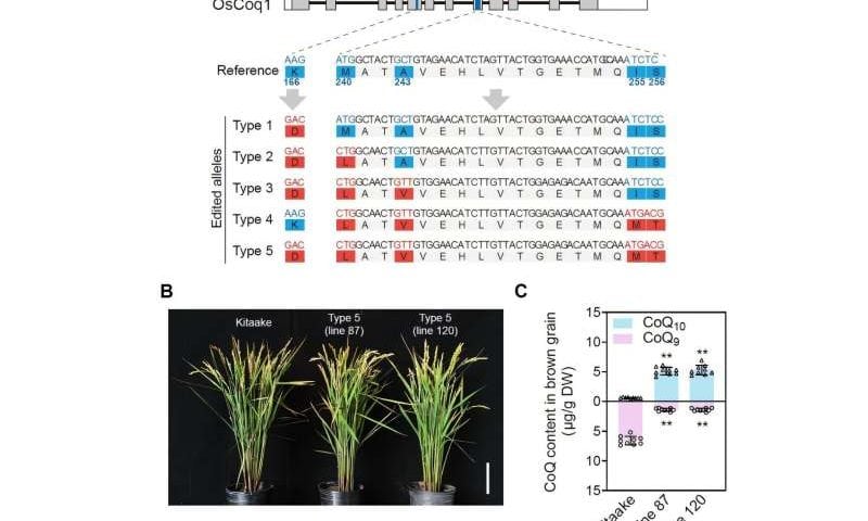
Led by Prof. Chen Xiaoya from the CAS Center for Excellence in Molecular Plant Sciences/Shanghai Chenshan Research Center and Prof. Gao Caixia from the Institute of Genetics and Developmental Biology of the Chinese Academy of Sciences (CAS), the researchers used targeted gene editing to modify just five amino acids of the Coq1 rice enzyme, creating new rice varieties capable of synthesizing CoQ10.

The study is published in Cell.

CoQ10 is important for human health, especially heart protection. It is an essential component of the mitochondrial electron transport chain and serves as a fat-soluble antioxidant. Different species synthesize CoQ with different side-chain lengths.

Humans synthesize CoQ10 with a side chain of 10 isoprene units (C50), while cereal food crops like rice and wheat, and certain vegetables and fruits, primarily synthesize CoQ9, which contains nine isoprene units (C45).

The invention of CoQ10-producing crops to substantially increase the CoQ10 content in plant-based foods offers a cost-effective and environmentally friendly approach to nutritional fortification, offering great potential benefit.

The molecular mechanism underlying the variation in CoQ side-chain length was previously unclear. Thanks to the diverse plant collections at the Shanghai Chenshan Botanical Garden of the CAS Center for Excellence in Molecular Plant Sciences, the research team obtained 134 plant samples from 67 families, including mosses, clubmosses, ferns, gymnosperms, and angiosperms.

The researchers determined the distribution patterns of CoQ types in these species and revealed that CoQ10 is an ancestral trait of flowering plants, with most plants still synthesizing CoQ10. However, grasses, daisies, and cucurbit plants mainly produce CoQ9.

After analyzing the evolutionary trajectories and natural variations of Coq1 enzymes across more than 1,000 terrestrial plant species, as well as by using machine learning, the researchers ultimately identified five amino acid sites that determine chain length.

After the targeted editing, the rice plants in this study primarily synthesized CoQ10, with up to 5 μg/g produced per rice grain. This shows that gene editing has become an efficient and safe technology for crop breeding.

The successful development of CoQ10 rice will significantly expand the food sources of CoQ10. It also provides an example of using big data and AI for crop breeding.

CRISPR system variant shows a novel DNA cleavage mechanism

Researchers have revealed new details about the CRISPR-Cas5-HNH/Cascade complex, a variant of the type I-E CRISPR-Cas system, providing insights into its DNA recognition and cleavage mechanisms.

The study, published in Nature Communications, was conducted by researchers from the Institute of Physics of the Chinese Academy of Sciences.

The CRISPR-Cas5-HNH/Cascade complex, which serves as an immune defense system in prokaryotes, has long been studied for its ability to protect bacteria from invading genetic materials. However, recent research uncovers a novel mechanism in this complex that differs significantly from the well-known type II CRISPR systems like Cas9.

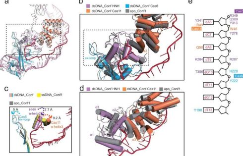
Researchers utilized cryo-electron microscopy to capture the structures of the Cas5-HNH/Cascade complex in both DNA-bound and unbound states. They revealed striking conformational changes. The complex adopts a more compact structure when bound to double-stranded DNA (dsDNA), with the target DNA strand making a pronounced U-turn and interacting with the HNH nuclease domain.

The target strand is cleaved first, followed by the non-target strand, a departure from the cleavage order seen in other CRISPR systems like Cas12a.

The Cas5-HNH domain itself was found to play a crucial role in the nuclease activity of the Cas5-HNH/Cascade complex. Researchers demonstrated that mutations in key residues of the HNH domain, particularly histidine and aspartate, can completely abolish cleavage activity.

This suggests that the HNH domain is not only essential for target DNA recognition but also for the specific cleavage of DNA, further differentiating the mechanism from other systems.

Moreover, researchers observed that the architecture of the Cas5-HNH/Cascade complex is notably different from canonical type I-E systems. For example, the HNH domain was fused to the C-terminus of Cas5, replacing the need for Cas3, a protein commonly involved in DNA degradation in other systems.

This structural innovation pointed to the complex’s ability to cleave DNA independently of the traditional components seen in similar CRISPR systems.

By elucidating the role of the HNH nuclease domain and revealing the structural changes that occur upon DNA binding, this work not only deepens the understanding of CRISPR-Cas5-HNH but also paves the way for refining CRISPR technologies for therapeutic applications.

Colon cancer among young people is on the rise. Could yogurt help?

Yogurt is not only just part of a delicious breakfast.

A new study led by Mass General Brigham researchers found it may also have protective benefits against a certain type of aggressive colon cancer.

Study authors analyzed data totaling over 150,000 people who answered questionnaires about lifestyle factors and disease outcomes, including questions about their intake of plain and flavored yogurt, as well as other dairy products.

These participants were followed for at least three decades.

Researchers found people who ate two or more servings of yogurt a week had lower rates of proximal colon cancer that were positive for Bifidobacterium, according to the study published Wednesday in the peer-reviewed journal Gut Microbes.

Proximal colon cancer is a type of colorectal cancer that occurs on the right side of the colon and has been shown to have worse survival outcomes compared to patients with distal cancers, which occur more on the left side.

“It has long been believed that yogurt and other fermented milk products are beneficial for gastrointestinal health,” said co-senior author Dr. Tomotaka Ugai, a pathology investigator at Brigham and Women’s Hospital. “Our new findings suggest that this protective effect may be specific for Bifidobacterium-positive tumors.”

Colorectal cancer is the third-leading cause of cancer-related deaths in men and the fourth-leading cause in women, according to the American Cancer Society.

The organization estimates nearly 53,000 people will die from it in 2025.

While the death rates from colorectal cancer have been dropping in older adults, rates have been increasing by about 1% per year since the mid-2000s in people under 55.

The new yogurt study adds to a growing body of evidence that shows the connection between diet, the gut microbiome and the risk of colorectal cancer, said co-author Dr. Andrew T. Chan, chief of clinical and translational epidemiology at Massachusetts General Hospital.

“It provides an additional avenue for us to investigate the specific role of these factors in the risk of colorectal cancer among young people,” he said.

error: Content is protected !!