Designing self-destructing bacteria to make effective tuberculosis vaccines

Working toward more effective tuberculosis (TB) vaccines, researchers at Weill Cornell Medicine have developed two strains of mycobacteria with “kill switches” that can be triggered to stop the bacteria after they activate an immune response. Two preclinical studies, published Jan. 10 in Nature Microbiology, tackle the challenge of engineering bacteria that are safe for use in controlled human infection trials or as better vaccines. While TB is under control in most developed countries, the disease still kills over a million people a year worldwide.

Spreading easily through the air, Mycobacterium tuberculosis can establish a chronic infection in human lungs, which can turn into a deadly respiratory disease. A safe vaccine called BCG, consisting of a weakened strain of the closely related Mycobacterium bovis, has been available for over a century but has limited efficacy.

“BCG protects children from tuberculosis meningitis, but it doesn’t effectively protect adults from pulmonary tuberculosis, which is why it’s only used in high-incidence countries,” said Dr. Dirk Schnappinger, professor of microbiology and immunology at Weill Cornell Medicine and a senior author on both of the new studies.

However, collaborators at the University of Pittsburgh and the National Institutes of Health’s Vaccine Research Center previously found that administering high doses of the BCG vaccine directly into the veins, instead of the usual route of giving it under the skin, was better at protecting adult macaque monkeys against lung infection.

Building a better vaccine

In one of the new papers, the team aimed to make this high-dose intravenous injection safer, without destroying the vaccine’s ability to stimulate a strong immune response. “We needed a version of BCG that triggers an immune response, but then you can flip a switch to eliminate the bacteria,” said Dr. Schnappinger.

After testing about 20 different strategies, the investigators found that lysins, enzymes encoded by viruses that can infect BCG, cause the bacteria to self-destruct. Using a clever bit of molecular engineering, they placed two different lysin genes under the control of gene regulators that respond to an antibiotic. By adding or taking away the antibiotic, they could then flip the kill switch.

“The lysins were known, but I don’t think they have been utilized as kill switches previously,” said Dr. Sabine Ehrt, professor of microbiology and immunology at Weill Cornell Medicine and a senior author on the papers.

With the newly engineered BCG, the researchers delivered high doses of the vaccine intravenously to antibiotic-treated macaques. When they stopped the antibiotic, the kill switch was activated, promptly ending the infection. The self-destructing bacteria released antigens that further stimulated the animals’ immune systems. The result was a robust immune response that protected the monkeys from subsequent lung infections with M. tuberculosis.

“Despite the promising preclinical results, evaluating if the vaccination actually works takes a long time and many people to test it. Tuberculosis doesn’t develop quickly and only in a small fraction of the people who are infected,” Dr. Schnappinger explained.

Such enormous, lengthy clinical trials can cost hundreds of millions of dollars, a major barrier to new vaccines. The urgent need for an effective TB vaccine has prompted researchers to find innovative ways to accelerate vaccine development.

3D imaging approach reveals intricate steps of herpes simplex virus assembly

A new combination of microscopy methods has revealed exquisite detail of the virus assembly process used by herpes simplex virus during replication.

The research, published today in eLife, is described by the editors as a fundamental study that comprehensively examines the roles of nine structural proteins in herpes simplex virus 1 (HSV-1) viral assembly. They say the thoroughly executed research yields compelling data that explain previously unknown functions of HSV-1 structural proteins.

Additionally, by integrating cryo-light microscopy and soft X-ray tomography, it presents an innovative approach to investigating viral assembly within cells that will be of broad interest to virologists, cellular biologists and structural biologists.

HSV-1 is a large virus that infects the mucous membranes of the mouth and genitals, causing life-long latent infections. The virus is composed of three layers—a capsid that contains the viral DNA, a protein layer called the tegument and an outer envelope that is studded with viral glycoproteins (proteins with a sugar attached). During replication, newly copied viral genomes are packaged up into this three-layer structure in a process called viral assembly.

While some drugs can block the virus’ DNA replication and alleviate symptoms, there is no permanent cure. A deeper understanding of the assembly process could inform the design of novel treatments or cures that inhibit virus formation.. But until now, pinpointing the role of different HSV-1 components in the viral assembly process has proved challenging.

“HSV-1 mutants that cannot make certain proteins have been used to study the role of viral genes in virus assembly, using a method called thin section transmission electron microscopy, or TEM,” says lead author Kamal Nahas, Beamline Scientist at Beamline B24, Diamond Light Source, Harwell Science & Innovation Campus, Didcot, U.K. “However, the extensive sample processing required for TEM can distort the microscopic structure and complicate the interpretation of features in viral assembly.”

Viral assembly involves a multi-step process, starting in the cell’s nucleus with assembly of capsids, packaging of the DNA to form “nucleocapsids,” and transport of these nucleocapsids out of the nucleus via a process of primary envelopment and de-envelopment to travel across the nuclear envelope. This is followed by a secondary envelopment in the cellular area surrounding the nucleus, called the cytoplasm (cytoplasmic envelopment).

Imaging methods that maintain the HSV-1-infected cells as close to physiological conditions as possible are needed to fully understand this complex, three-dimensional (3D) assembly process.

The authors used an emerging 3D imaging approach to study the envelopment mechanism and investigate the importance of different HSV-1 genes for viral assembly by investigating the impact of specific mutations of these viral genes. Their new approach combined two methods—cryo-structured illumination microscopy (cryoSIM) to detect fluorescently labeled capsid or envelope components, and cryo-soft-X-ray tomography (cryoSXT) to identify the cellular substructure in the same infected cells.

Together, this “correlative light X-ray tomography” (CLXT) approach makes it possible to identify specific structural components within the viral assembly process, allowing the team to visualize exactly where the assembly process stalls for each mutant virus, and providing insights into the unmutated gene’s usual role in viral assembly.

The authors captured different assembly stages during cytoplasmic envelopment using their mutant viruses and showed that—contrary to previous theories—cytoplasmic envelopment is caused by the budding of a capsid into an intracellular membrane “sack” or vesicle, and not by the capsid being “wrapped” by the vesicle membrane.

A further new finding is that this budding is asymmetric; the team observed several instances of stalled viral assembly where groups of capsids were gathered at one region, or side, of a spherical vesicle.

Using their CLXT approach, they were able to rank the relative importance of five of the mutant viral proteins in the process of nuclear egress. They were also able to reveal the role of a further five viral proteins in the cytoplasmic envelopment stage. For example, a protein called VP16 was found to be important in delivering the capsid to envelopment compartments and is now thought to have a larger role in nuclear egress than previously thought. In addition, the new method revealed that the absence of four other proteins caused virus particles to build up in the cytoplasm where assembly had stalled.

“Our multi-modal imaging strategy has provided novel ultrastructural insight into HSV-1 assembly, allowing the assembly trajectory of normal and mutant viruses to be observed in 3D,” concludes senior author Colin Crump, Professor of Molecular Virology at the University of Cambridge, U.K. “Our data underscore the power of correlative fluorescence and X-ray tomography cryo-imaging for interrogating and conducting further studies on the process of virus assembly.”

Think five minutes isn’t enough time to make a difference in terms of health and well-being?

It could actually be enough time to stave off a disease that afflicts many people in their later years.

Just five minutes of light exercise a day could help prevent dementia, even for frail older adults, new research has found.

Dementia Risk May Be Lowered By One Important Medical Device

The latest study on the topic was led by researchers at the Johns Hopkins Bloomberg School of Public Health in Baltimore, Maryland.

They found that engaging in as little as 35 minutes of moderate to vigorous physical activity per week — as compared to none at all — was associated with a 41% lower risk of developing dementia over an average four-year follow-up period.

The findings were published recently in The Journal of the American Medical Directors Association and shared on several medical sites.

Even for people at an elevated risk of “adverse health outcomes,” greater activity was associated with lower dementia risk, the researchers noted.

The higher amounts of physical activity, the lower the risk of dementia.

Consider this data from the study: Dementia risks were 60% lower in participants who got 35-to-69.9 minutes of physical activity/week; 63% lower in the 70-to-139.9 minutes/week category; and 69% lower in the 140-and-over minutes/week category.

For their analysis, the researchers analyzed a dataset covering nearly 90,000 adults living in the United Kingdom who wore smartwatch-type activity trackers, news agency SWNS reported.

Lead study author Dr. Amal Wanigatunga said, “Our findings suggest that increasing physical activity, even as little as five minutes per day, can reduce dementia risk in older adults.”

Dementia, usually in the form of Alzheimer’s, affects millions of people all over the globe.

While public health guidelines usually recommend 150 minutes of moderate activity per week, the study aligns with a growing body of evidence supporting a “some-is-better-than-none” approach to physical activity, according to Study Finds.

And while the risk of Alzheimer’s increases with age, recent research has suggested it may be somewhat preventable by certain lifestyle changes, including better control of cholesterol, blood pressure and blood sugar — plus being more active, SWNS noted.

Participants in the new study had a median age of 63. Women made up 56% of the sample.

Over an average follow-up period of 4.4 years, 735 people among the group developed dementia.

Researchers found that for every additional 30 minutes of weekly moderate to vigorous physical activity (MVPA), there was a 4% reduction in dementia risk.

But the most “striking” finding came when comparing people who engaged in no physical activity at all to those who managed to get even minimal amounts.

“This suggests that even frail or nearly frail older adults might be able to reduce their dementia risk through low-dose exercise,” said Wanigatunga.

He noted that the study was not a clinical trial that established causation indicating that exercise reduces dementia risk, but that its findings are consistent with that hypothesis.

“To check the possibility that their findings reflected undiagnosed dementia leading to lower physical activity,” News Medical reported, “the researchers repeated their analysis but excluded dementia diagnoses in the first two years of follow-up.”

“The association between more activity and lower dementia risk remained robust.”

Dr. Marc Siegel, clinical professor of medicine at NYU Langone Health and Fox News’ senior medical analyst, was not involved in the study but shared his reaction to the “important” findings.

“This is not proof, just an association, but is very useful for the group that gets discouraged, thinking, ‘I can’t do a lot of exercise because of illness or disability, so why do any?’ This study suggests that even small amounts are helpful.”

There are many mechanisms that could explain this effect, Siegel said – “primarily increased blood flow to the brain, as well as improved disposal of metabolic waste and decreased inflammation.”

He added, “It is also likely associated with healthier lifestyle decisions that also decrease the advent of neuroinflammation, dysregulation and plaque formation that characterize dementia, especially Alzheimer’s.”

Some of these include sleep, diet and engagement, he said.

The National Institute on Aging provided funding for the new study.

New coronavirus found in bats is not currently ‘concern to public health’: CDC

The Centers for Disease Control and Prevention (CDC) said on Monday that the new coronavirus found in bats is currently not a cause for concern.

There is no reason to believe the virus poses a threat to public health at the moment and no infections have been detected in humans, according to the federal health agency.

“CDC is aware of a publication about a new bat coronavirus, but there is no reason to believe it currently poses a concern to public health,” the agency said in a statement. “The publication referenced demonstrates that the bat virus can use a human protein to enter cells in the laboratory, but they have not detected infections in humans.”

Chinese researchers, including from the Wuhan Institute of Virology and Guangzhou Medical University, published a paper in the journal Cell on Friday indicating they had discovered a new bat coronavirus that could have the potential to infect humans.

The newer coronavirus is known as HKU5-CoV-2 and is a type of merbecovirus, which is the same family of another coronavirus known to infect humans called Middle East Respiratory Syndrome (MERS).

In a lab study, the new coronavirus was found to have the potential to enter cells through the ACE2 receptor, a protein found on the cells’ surface.

This is the same way the virus that causes COVID-19 infects people, which theoretically means the new coronavirus could pose a risk to spilling over into humans.

The spike protein of the new coronavirus infected human cells that had high levels of the ACE2 receptor in test tubes, as well as in small models of human airways and intestines.

The researchers found that the virus did not enter human cells as readily as the virus that causes COVID-19 — which is called SARS-CoV-2 — writing that the “risk of emergence in human populations should not be exaggerated.”

None of the animal studies that were conducted examined the virus’s ability to cause disease or its transmissibility.

If the virus were to infect humans, the researchers suggested antiviral drugs and monoclonal antibodies — laboratory-produced proteins that mimic the antibodies the body naturally creates when fighting a virus — could be effective.

There are hundreds of coronaviruses circulating in nature. Only a few can infect humans, causing illnesses ranging from mild respiratory tract infections to more severe conditions such as bronchitis or pneumonia.

Coronaviruses include some variations of the common cold, the virus that causes MERS, severe acute respiratory syndrome (SARS) and the virus that causes COVID-19.

The researchers wrote that “bats harbor the highest proportion of genetically diverse coronaviruses,” posing a risk of spilling over into humans.

Genome-edited rice shows resistance to bacterial blight in East Africa

The international Healthy Crops consortium has developed an innovative strategy to combat the disease bacterial blight (for short: BB) in rice using genome editing technology. If approved for use by farmers in Kenya, the BB-resistant rice varieties are expected to reduce yield losses associated with the disease in the affected rice growing regions and increase productivity. The work is a collaboration between Kenya Agricultural and Livestock Research Organization (KALRO) and Heinrich Heine University Düsseldorf (HHU).

Rice production is of central importance for food security and economic development in many countries, in particular in low- and medium-income countries. Rice is the second most important staple food in East Africa, with 1.8 million tonnes consumed every year in the countries of the East African Community (for short: EAC).

In 2019, members of the Healthy Crops team identified an outbreak of BB in Tanzania caused by invasive Asian variants of the bacterium Xanthomonas oryzae pv. oryzae (Xoo). The bacterium is spreading rapidly and causing estimated yield losses of 13–20%.

Dr. Emily Gichuhi from KALRO explains, “Due to climate change, incidences of rice diseases including BB have been on the rise in Kenyan rice growing areas. This has increased the cost of production among rice farmers, thereby reducing their returns.”

Dr. Daigo Makihara and Dr. Moto Ashikari from Nagoya University (NU) in Japan, researchers from the Wonder Rice Initiative for Food Security and Health (WISH), are working closely with Dr. Gichuhi and her team to develop new African rice varieties. Dr. Makihara explains, “As a result of the international spread of different crop plant varieties, we are increasingly finding ourselves confronted with outbreaks of plant diseases in regions where they have not previously played a role.”

The starting point for the researchers from Healthy Crops is the nutrient supply of the bacteria. The Xoo bacteria possess a set of “keys” which can open the “pantry” of the plants: When the bacterium injects one of these “key” proteins into rice cells, it leads to increased production of a transporter, which releases sugar in the neighborhood of the bacteria. This sugar serves as nutrition and is essential for the multiplication and virulence of the bacteria. However, when the bacteria utilize the sugar, there is none left for the plant, which ultimately dies as a result

The research team has succeeded in changing the “locks” via genome editing, making the plants resistant to all known Xoo strains currently prevalent in Asia and Africa.

Professor Bing Yang, University of Missouri, who developed the editing approach, states: “The combination of two different sets of enzymes for editing enabled us to develop a robust resistance.”

The import of these edited elite rice varieties has been made possible due to the availability of genome editing guidelines developed by the National Biosafety Authority (NBA) of Kenya and published in 2022.

Dr. Marcel Buchholzer, coordinator of the Healthy Crops project at HHU, explains, “It is now possible to evaluate these rice lines, developed using advanced biotechnology methods at HHU, in Kenya.”

Professor Dr. Wolf B. Frommer, spokesperson for the project at HHU explains, “This project aims to protect smallholder farmers from crop yield losses through knowledge-based approaches to fighting plant diseases.”

Circular mRNA produces 200 times more protein, enhancing precision therapy potential

Imagine a breakthrough in cancer treatment where only malignant cells are targeted, sparing healthy host cells; or patients with abnormal protein synthesis are treated to produce a healthy protein. Hiroshi Abe and his colleagues at Nagoya University have identified two applications, among others, in a new study.

Their innovative approach, reported in Nature Biotechnology, called the Internal Cap-Initiated Translation (ICIT) mechanism, introduces a novel way to “switch on” protein synthesis only in target cells, creating healthy proteins to treat illnesses or toxic proteins to kill unwanted cells.

Capping circular mRNA in a new way

ICIT builds on the promise of circular mRNAs, a new generation of mRNA treatments known for their stability and reduced inflammatory effects compared to traditional linear mRNAs.

Unlike linear mRNAs, circular mRNAs are less susceptible to enzymatic degradation because of their lack of terminal structures, offering a sustained translation process.

However, one significant challenge with circular mRNAs has been the inefficiency of their translation inside living organisms.

Previous methods relied on long internal ribosome entry sites (IRES) for introducing the mRNA, which were difficult to optimize and often inefficient. Abe’s team overcame this hurdle by introducing a cap structure into the circular mRNA itself.

This internal cap structure triggers translation initiation, bypassing the need for IRES sequences, and significantly improves the efficiency of protein synthesis.

Precision therapy

Abe and his colleagues developed two designs. Among these, Cap-circRNA demonstrated superior performance, synthesizing up to 200 times more protein than commonly used circular mRNAs with IRES sequences. Importantly, this synthesis persisted for an extended period, even after traditional mRNA structures began to degrade.

This stability and ability to selectively target cells make Cap-circRNA an ideal candidate for developing precision therapies.

“This technology is expected to revolutionize mRNA medicine, including antibody therapy, genome editing, and protein replacement therapy,” Abe said.

“Current mRNA is fundamentally unstable, requiring constant injections to be used for treatments such as protein replacement, a problem that our technique overcomes. Using this, we could treat diseases caused by abnormal protein synthesis, such as Duchenne muscular dystrophy.”

Targeting cancer cells

The ICIT mechanism’s ability to control protein translation at the single-cell level also offers a transformative approach to the treatment of cancers and other tissue-specific diseases. By targeting specific RNA markers that are highly expressed in diseased cells, such as those found in liver cancer, the mRNA can instruct protein synthesis only in target cells.

This precision reduces the risk of off-target effects and side reactions, which are common challenges in current treatments. To test its efficacy, the team designed a circular RNA using ICIT to target HULC lncRNA, an RNA that is commonly found in liver cancer cells.

AI Model Measures Pace of Brain Aging, Could Aid Prediction of Cognitive Decline

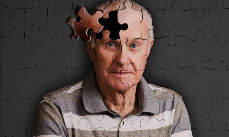
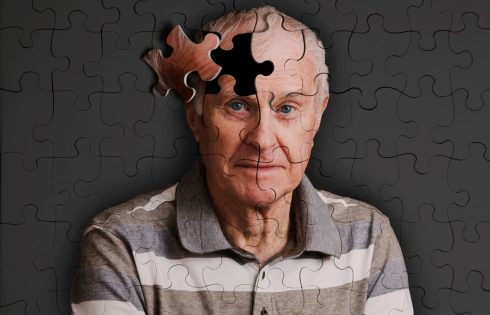
Scientists at the University of Southern California (USC) have developed an artificial intelligence (AI) model that they say could help scientists better understand, prevent, and treat cognitive decline and dementia.

The first-of-its-kind three-dimensional convolutional neural network (3D-CNN) tool noninvasively analyses magnetic resonance imaging (MRI) scans from an individual patient to track brain changes with time and measure the pace—P—of brain aging.

Faster brain aging closely correlates with a higher risk of cognitive impairment, said Andrei Irimia, PhD, associate professor of gerontology, biomedical engineering, quantitative & computational biology, and neuroscience at the USC Leonard Davis School of Gerontology and visiting associate professor of psychological medicine at King’s College London. “This is a novel measurement that could change the way we track brain health both in the research lab and in the clinic. Knowing how fast one’s brain is aging can be powerful.”

Irimia is the senior author of the study, published in the Proceedings of the National Academy of Sciences, that describes the new model and its predictive power. In the team’s report, titled “Deep learning to quantify the pace of brain aging in relation to neurocognitive changes,” Irima and colleagues concluded, “This research complements existing strategies for AD risk assessment that estimate individuals’ rates of adverse cognitive change with age.”

Biological age (BA) is distinct from an individual’s chronological age (CA), Irimia said. Two people who are the same age based on their birthdate can have very different biological ages due to how well their body is functioning and how “old” the body’s tissues appear to be at a cellular level. “Mapping the pace P of brain aging can help to identify abnormal rates of neural aging that may reflect neurodegenerative disease risk,” the team stated. “Whereas neurodegenerative disease risk increases with chronological age (CA), biological aging varies across cells, tissues, organs, and individuals.”

Some common measures of biological age use blood samples to measure epigenetic aging and DNA methylation, which influences the roles of genes in the cell. However, measuring biological age from blood samples is a poor strategy for measuring the brain’s age.

The barrier between the brain and the bloodstream prevents blood cells from crossing into the brain, such that a blood sample from one’s arm does not directly reflect methylation and other aging-related processes in the brain Conversely, taking a sample directly from a patient’s brain is a much more invasive procedure, making it unfeasible to measure DNA methylation and other aspects of brain aging directly from living human brain cells. “Measuring P is challenging due to its dynamic nature throughout life,” the authors wrote. “The pace of aging is frequently estimated based on DNA methylation of whole-blood cells. However, this is not ideal for the brain because the blood–brain barrier separates neural cells from the blood physically and biochemically.”

Previous research by Irimia and colleagues highlighted the potential of MRI scans to non-invasively measure the biological age of the brain. The earlier model used AI analysis to compare a patient’s brain anatomy to data compiled from the MRI scans of thousands of people of various ages and cognitive health outcomes.

However, the cross-sectional nature of analyzing one MRI scan to estimate brain age had major limitations. While the previous model could, for instance, tell if a patient’s brain was ten years “older” than their calendar age, it couldn’t provide info on whether that additional aging occurred earlier or later in their life, nor could it indicate whether brain aging was speeding up.

Created in collaboration with Paul Bogdan, PhD, associate professor of electrical and computer engineering and holder of the Jack Munushian Early Career Chair at the USC Viterbi School of Engineering, the newly developed 3D-CNN offers a more precise way to measure how the brain ages over time, by analyzing MRI scans taken at different time points for the same patient. Unlike traditional cross-sectional approaches, which estimate brain age from one scan at a single time point, the new longitudinal model (LM) compares baseline and follow-up MRI scans from the same individual. As a result, it more accurately pinpoints neuroanatomic changes tied to accelerated or decelerated aging.

The authors first trained and validated the model on more than 3,000 MRI scans of cognitively normal (CN) adults. When applied to a group of 104 cognitively healthy adults and 140 Alzheimer’s disease patients, the new model’s calculations of brain aging speed closely correlated with changes in cognitive function tests given at both time points. The 3D-CNN also generates interpretable “saliency maps,” which indicate the specific brain regions that are most important for determining the pace of aging, Bogdan said. “The alignment of these measures with cognitive test results indicates that the framework may serve as an early biomarker of neurocognitive decline. “Moreover, it demonstrates its applicability in both cognitively normal individuals and those with cognitive impairment.”

Bogdan further commented that the model has the potential to better characterize both healthy aging and disease trajectories, and its predictive power could one day be applied to assessing which treatments would be more effective based on individual characteristics. “Estimated P values correlate significantly with changes in cognitive function, suggesting its utility for monitoring abnormal brain aging rates during neurodegeneration,” the scientists stated. “Rates of brain aging are correlated significantly with changes in cognitive function,” Irimia noted. “So, if you have a high rate of brain aging, you’re more likely to have a high rate of degradation in cognitive function, including memory, executive speed, executive function, and processing speed. It’s not only an anatomic measure; the changes we see in the anatomy are associated with changes we see in the cognition of these individuals.”

In their study, Irimia and coauthors noted how the new model was able to distinguish different rates of aging across various regions of the brain. Delving into these differences—including how they vary based on genetics, environment, and lifestyle factors—could provide insight into how different pathologies develop in the brain, Irimia said.

The study also demonstrated that the pace of brain aging in certain regions differed between the sexes, which might shed light on why men and women face different risks for neurodegenerative disorders, including Alzheimer’s, he added. “By synergizing the LM with an interpretable CNN saliency approach, we map anatomic variations in regional brain aging rates that differ according to sex, decade of life, and neurocognitive status,” the investigators stated. “LM estimates of P are significantly associated with changes in cognitive functioning across domains. This underscores the LM’s ability to estimate P in a way that captures the relationship between neuroanatomic and neurocognitive aging.”

Irimia said he is also excited about the potential for the new model to identify people with faster-than-normal brain aging before they show any symptoms of cognitive impairment. While new drugs targeting Alzheimer’s have been introduced, their efficacy has been less than researchers and doctors have hoped for, potentially because patients might not be starting the drug until there is already a great deal of Alzheimer’s pathology present in the brain, he explained. “Individually tailored strategies to reduce P could increase healthspan and maintain functions that diminish with age,” the team concluded.

“One thing that my lab is very interested in is estimating risk for Alzheimer’s; we’d like to one day be able to say, ‘Right now, it looks like this person has a 30% risk for Alzheimer’s,’” Irima said. “We’re not there yet, but we’re working on it. I think this kind of measure will be very helpful to produce variables that are prognostic and can help to forecast Alzheimer’s risk. That would be really powerful, especially as we start developing potential drugs for prevention.”

Study Shows Anti-Aging Potential for Insilico’s IPF Candidate

Researchers from artificial intelligence (AI) drug developer Insilico Medicine and two partner institutions have published a study concluding that its lead candidate ISM001-055 is the first to show anti-aging properties and that its mechanism of action offers a promising therapeutic approach for treating age-related diseases.

ISM001-055 was shown to have attenuated cellular senescence through the suppression of various aging processes, thus showing potential as a senomorphic drug. Senomorphics are a class of drugs that targets the senescence-associated secretory phenotype (SASP) of senescent cells, which have stopped dividing and accumulate in tissues as people age, thus are considered to play a role in aging and age-related diseases.

The study also showed ISM001-055 to function as a senomorphic agent, modulating the behavior of senescent cells rather than eliminating them. ‘055 not only matched, but also surpassed the FDA-approved sirolimus, formerly rapamycin—sold by Pfizer as Rapamune® and by several other companies as generic versions—in two key areas:

  • Reduction of SASP factors: ISM001-055 was more effective than rapamycin in decreasing the secretion of pro-inflammatory cytokines and other SASP components, which are linked to tissue dysfunction and age-related pathologies.
    • Restoration of cellular function: Cells treated with ISM001-055 exhibited a more pronounced return to youthful functionality compared to those treated with rapamycin, indicating superior rejuvenative properties.

    ISM001-055 is an internally developed Insilico drug candidate developed using generative AI. The drug is designed to treat idiopathic pulmonary fibrosis (IPF) by targeting Traf2- and NCK-interacting kinase (TNIK), a serine/threonine kinase whose activation plays a crucial role in cellular processes that include signal transduction pathways essential for fibrosis development.

    In inhibiting TNIK, ISM001-055 reduced the activation of the TGF-β and Wnt/β-catenin pathways, which are involved in SASP regulation. This enabled ‘055 to suppress pro-inflammatory cytokine production while preserving senescent cells that may still serve beneficial functions, such as in tissue repair and tumor suppression.

    “The strong evidence that TNIK plays a role in aging and senescence could influence Insilico to expand the development of ISM001-055 beyond fibrosis and into broader geroprotective and anti-aging applications,” Insilico founder and CEO Alex Zhavoronkov, PhD, told GEN Edge.

    Insilico’s future plans for INSM001-055 are undisclosed.

    Implicating TNIK in aging

    “This study further strengthens the potential of INS018_055 as a longevity therapeutic by implicating TNIK in the cellular senescence hallmark of aging,” the researchers concluded, using the drug candidate’s former name, in “AI-Driven Robotics Laboratory Identifies Pharmacological TNIK Inhibition as a Potent Senomorphic Agent,” a study published in Aging and Disease. The Insilico researchers were joined by investigators from China’s Suzhou Hospital of Nanjing Medical University, as well as from the Buck Institute for Research on Aging.

    “This research highlights the novel role of TNIK in cellular senescence and new senomorphic applications for INS018_055, in addition to its anti-fibrotic properties that may inform future efforts to treat age-related diseases,” the researchers added.

    ISM001-055 has generated positive results in a “Phase 0” microdose trial (ACTRN12621001541897) and two Phase I clinical trials, one conducted in New Zealand (NCT05154240) and the other, in China (CTR20221542)—as well as a Phase IIa trial conducted across 21 sites in China (NCT05938920). That study’s secondary efficacy endpoint showed dose-dependent improvements in forced vital capacity (FVC), with the largest improvement observed in the 60 mg QD [once daily] cohort.

    Insilico is working to validate findings from these studies through a parallel Phase IIa trial (NCT05975983) now enrolling patients in the United States. The study is projected to enroll a total of 60 patients and achieve primary completion in February 2026.

    However, the results detailed in the latest paper came not from these clinical trials but from a study Insilico carried out at its AI-based, sixth-generation robotics laboratory in Suzhou, China, which according to the company, allowed for increased validation and consistency across experiments.

    “Insilico conducted this separate study using its AI-driven robotics lab to explore the senomorphic potential of ISM001-055 beyond its known anti-fibrotic effects,” Zhavoronkov said. “The robotic study provides high-throughput, automated, and AI-driven analysis of cellular responses, allowing for a detailed mechanistic understanding of how ISM001-055 impacts cellular senescence. This approach enables the identification of molecular pathways, aging biomarkers, and transcriptional changes in response to treatment.”

    Reducing inflammatory cytokines

    Researchers found that ISM001-055 primarily attenuates SASP by reducing inflammatory cytokines such as IL-6, IL-8, IL-1A, and IL-1B. ‘055 also attenuates extracellular matrix (ECM) remodeling, preventing excessive fibrosis and tissue dysfunction; as well as TGF-β signaling, a major driver of fibrosis and senescence-associated inflammation.

    In addition to reducing inflammation, results of attenuation included improving mitochondrial function, and outright increasing of healthy years or “healthspan.”

    Which effects is Insilico most interested in seeing?

    “Improving mitochondrial function and increasing healthspan are incredibly important and a major goal,” Zhavoronkov explained. “The most immediate translational impact comes from mitigating chronic inflammation and ECM remodeling, which are major drivers of aging-related diseases.”

    Zhavoronkov added that Insilico will continue focusing on reducing inflammation and SASP, since they contribute directly to multiple age-related diseases, including fibrosis and chronic inflammatory conditions.

    “Insilico will continue to confirm the findings by conducting more validating studies to ensure that the senomorphic effects observed in cellular models translate into whole-organism benefits. As progress continues, additional trials targeting aging-related diseases and conditions driven by senescence may be launched,” Zhavoronkov said.

    The researchers acknowledged they will need to collect additional data to support ISM001-055’s use for anti-aging therapy in clinical settings. According to Zhavoronkov, potential ways to further collect this data may include:

    • Animal model studies intended to validate ‘055’s effects on senescence and aging in vivo
    • Biomarker studies designed to identify changes in aging-related molecular markers in treated organisms
    • Potential expansion of clinical trials to evaluate its effects on age-related diseases beyond IPF
    • AI-driven modeling to refine target selection and optimize dosing strategies for anti-aging effects

    In a paper published in March in Nature Biotechnology, a team of 30 researchers led by Zhavoronkov detailed how they used generative AI to discover INS018_055, with a novel target discovered by Insilico’s target identification engine, PandaOmics, and a novel molecular structure designed by its generative chemistry engine, Chemistry42. Both are specific-function platforms within the company’s AI platform, Pharma.AI.

    ISM001-055 is Insilico’s first wholly owned program in which AI was used to identify a novel target and generate novel small molecules through Pharma.AI. Insilico won the FDA’s first Orphan Drug Designation for an AI drug in 2023.

Utah’s attorney general targets safety issues over weight loss drugs

SALT LAKE CITY — Utah’s Attorney General Derek Brown signed onto a letter along with attorneys general from 37 other states to ask the Food and Drug Administration to enforce the safety of weight loss drugs.

The letter asks the FDA to “ramp up enforcement against any compounding pharmacies that may be illegally participating in the market,” per a press release issued Wednesday by Brown’s office.

Brown said Friday that because of our “physical location,” Utah is dealing with a drug epidemic that includes the danger of buying and selling fake drugs. He added in the press release that the FDA needs to further investigate and protect consumers “against all the illegal and deceptive conduct.”

Weight loss drugs like Ozempic or Wegovy have become popular in recent years. Their main ingredient, semaglutide, has been traditionally used to treat diabetics, but according to a study by KFF last year, 1 in 8 Americans have used it to lose weight.

Its popularity has caused people to scour the internet looking for the drug — or a cheaper knockoff.

“People are really taking a risk by ordering these products online and not knowing the supplier that’s sending it to them,” Salvatore Ingrassia, a port director for Customs and Border Protection at New York’s JFK Airport, told CBS News.

Although they may look legitimate, Ingrassia added, “We’ve found things like antifreeze and incorrect amounts of active ingredients in these products.”

The purple issues

Though he is new to the office, Brown told the Deseret News that he is the newest member of the executive committee of the National Association of Attorneys General.

Unlike the Republican and Democratic AG associations, Brown said the NAAG includes all 56 AGs — from 50 states and six U.S. territories — collaborating on the office’s “nuts and bolts and business” through a bipartisan lens.

He said the role involves establishing relationships with fellow attorneys, evaluating funding for critical programs and collaboratively addressing challenges that impact all communities.

“This is an opportunity for Utah to really work with and get to know all the other attorneys general in the country, help them understand what matters to us, and work within a bipartisan fashion,” Brown said. “There are so many issues where it doesn’t matter if it’s a red state or blue state; it matters.”

According to Brown, NAAG is tackling many pivotal issues aimed at enhancing the lives of Americans through legal means.

Not all generic drugs have the same quality, study finds

Generic prescription drugs usually save consumers money because they are the same as the name brand, but the patent has expired. But a new study published in the journal Production and Operations Management suggests not all generic drugs are the same.

Researchers say they have uncovered significant disparities in the safety of generic drugs manufactured in India compared to those produced in the United States. The research, led by In Joon Noh from Korea University, found that generic drugs made in India are linked to a 54% increase in severe adverse events, including hospitalization, disability, and death, compared to their U.S. counterparts.

The study, which involved matching 2,443 generic drugs from the U.S. and emerging economies, highlights the critical role of manufacturing location in drug safety. John Gray, co-author and professor at Ohio State University, said that regulatory and quality assurance practices differ significantly between emerging economies like India and advanced economies such as the United States. This difference, he noted, can have a substantial impact on drug safety.

A focus on India

Despite the FDA’s assurances that all generics should be equivalently safe and effective, the study’s findings challenge this notion, particularly concerning Indian-manufactured generics. George Ball, a co-author from Indiana University, stressed the importance of transparency in drug manufacturing locations to enhance consumer safety.

The research team, which includes experts who have collaborated with the FDA, were able to link drugs to their manufacturing plants using the Structured Product Labeling dataset, overcoming the FDA’s lack of transparency on this front. This breakthrough allowed for a direct comparison of pharmaceutically equivalent drugs from both countries.

The study also revealed that mature generic drugs, those on the market for a longer time, were primarily responsible for the increased adverse events. The researchers attributed this trend to intensified competition and cost-cutting measures in the pharmaceutical industry, which they suggested may compromise drug quality.

While the findings raise concerns, Gray cautioned against halting overseas production, acknowledging that there are both good and bad manufacturers in India and the U.S. Instead, he advocated for improved regulatory oversight, including unannounced inspections of overseas manufacturing plants, to ensure consistent drug quality.

The study’s authors recommend that the FDA increase transparency regarding drug manufacturing locations and quality, enabling consumers to make informed choices and incentivizing higher standards in the industry.

error: Content is protected !!