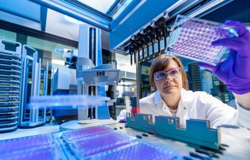
Emulsion technique boosts plant growth with bacteria

Researchers have demonstrated a technique for successfully encapsulating bacteria that can then be stored and applied to plants to improve plant growth and protect against pests and pathogens. The technique opens the door to creating a wide range of crop applications that allow farmers to make use of these beneficial bacteria in conjunction with agrochemicals. The paper, “Pickering Emulsion for Enhanced Viability of Plant Growth Promoting Bacteria and Combined Delivery of Agrochemicals and Biologics,” is published in the journal Advanced Functional Materials.

“Many of the beneficial bacteria we know of are fairly fragile, making it difficult to incorporate them into practical, shelf-stable products that can be applied to plant roots or leaves,” says John Cheadle, co-lead author of a paper on the work and a Ph.D. student at North Carolina State University. “The technique we demonstrate here essentially stabilizes these bacteria, making it possible to develop customized probiotics for plants.”

At issue are plant growth-promoting bacteria (PGPBs), which are microbes that benefit plant health and growth, helping plants extract nutrients from the environment and protecting them from pests or pathogens.

“A longstanding challenge for making use of these bacteria has been that if you tried to come up with a single application that combined them with agrochemicals, like pesticides or fertilizers, the bacteria would die,” says Saad Khan, co-corresponding author of the paper and INVISTA Professor of Chemical and Biomolecular Engineering at NC State. “We wanted to develop a solution that would allow bacteria to be used in conjunction with chemicals already in widespread use by growers.”

“By the same token, a healthy plant microbiome allows the plants to make better use of nutrients available in the soil and more resistant to pathogens,” says Tahira Pirzada, co-corresponding author and a research scholar at NC State. “This may allow growers to use less fertilizer and pesticides without hurting crop production.”

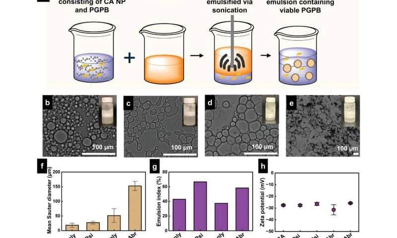
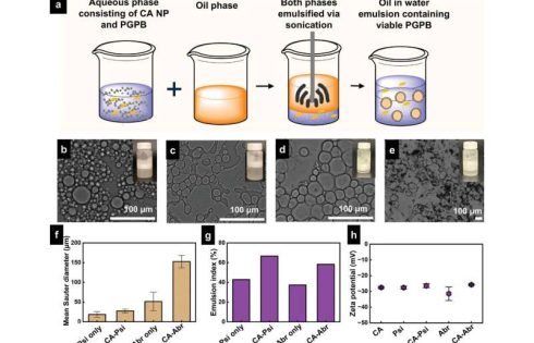
The new technique revolves around a custom-made emulsion, with only a handful of ingredients. One part of the emulsion consists of a saline solution that contains PGPBs. For the proof-of-concept demonstration, the researchers used the bacteria Pseudomonas simiae and Azospirillum brasilense. P. simiae acts as a biopesticide by promoting pathogen resistance; A. brasilense acts as a biofertilizer by fixing nitrogen.

The second part of the emulsion consists of a biodegradable oil and a biodegradable polymer derived from cellulose. The polymer can be loaded with agrochemical active ingredients, which means the emulsion can incorporate these ingredients without relying on environmentally harmful organic solvents, which are typically used in pesticide formulations.

When the two parts of the emulsion are mixed together, the oil is broken into droplets that are distributed throughout the saline solution. The cellulose polymer sticks to the surface of these droplets, preventing the droplets from merging back together.

Essentially, the emulsion is a salad dressing with the oil droplets held in suspension throughout the saline solution. In practical terms, this would allow the PGPBs to be applied simultaneously with agrochemicals using the same emulsion.

To see how well the emulsion worked, the researchers did two tests.

First, the researchers compared the survival of PGPBs in the emulsion to the survival of PGPBs in the saline solution alone. Samples of each were stored at room temperature. After four weeks, the population of P. simiae in the emulsion was 200% higher than the population in saline; the population of A. brasilense in the emulsion was 500% higher.

Engineered E. coli could be used to produce biodegradable plastics

E. coli bacteria could be used to create biodegradable plastics, reports a paper published in Nature Chemical Biology. The engineered bacterial system described in the study may help in the production of plastics with desirable thermal and mechanical properties, using renewable resources, the authors suggest.

Global plastic production is estimated to have created about 400 million metric tons of plastic in 2022, mostly through petroleum-based chemical processes. Meanwhile, the microbial production of polymers has the potential to develop biodegradable alternatives in a more sustainable way.

It is well known that organisms can naturally synthesize polymers, such as DNA, RNA, cellulose and proteins. However, scientists have only recently focused on the use of microorganisms to synthesize polymers that can be used to manufacture plastics.

Researcher Sang Yup Lee and colleagues developed a process to produce poly(ester amide) (PEA) using a series of enzymes produced in E. coli; this process involves combining one or more of six amino acids with one or more hydroxy acids to create the polymer plastic. After further tests to optimize the process, Lee and colleagues used glucose as a key ingredient to produce the polymers within E. coli. They also investigated how the amount and structure of the different amino acids used affects the production and properties of the PEAs.

As a proof of concept, the authors produced about 55 grams per liter of a PEA in a large bioreactor, demonstrating that PEA production can be easily scaled up. They also tested the physical, thermal and mechanical properties of this PEA, and suggest that they are comparable to those of high-density polyethylene, one of the most widely used plastics, which indicates that PEAs could serve as a renewable alternative.

This method has several advantages over current chemical methods, such as providing easy access to a wide range of PEAs and enabling the sustainable production of polymers that could be used as plastics, the authors conclude.

Mallinckrodt, Endo Plan $6.7B Merger

Mallinckrodt and Endo said they will merge through a $6.7 billion cash and stock deal creating a combined company that will be intent on bouncing back from their troubled recent histories, stemming from their involvement in marketing opioid drugs.

The combined company will have a presence in branded and generic drugs across a variety of therapeutic areas, a significant controlled substances franchise, and a product portfolio across multiple delivery technologies, formulations, and dosage forms.

The Mallinckrodt-Endo combo will start out with pro forma 2025 revenue of $3.6 billion and pro forma 2025 adjusted earnings before interest, taxes, depreciation, and amortization (EBITDA) of $1.2 billion.

However, the combined company expects to generate at least $150 million of annual pre-tax run-rate operating cost savings or “synergies” by its third-year post-closing of the merger, of which half or approximately $75 million is expected within a year of the merger close. The synergies, according to Mallinckrodt and Endo, will consist of business function integration and R&D savings from economies of scale, “among other areas.”

The Mallinckrodt-Endo combination will have approximately 5,700 employees at closing, as well as 17 manufacturing facilities, 30 distribution centers, and an operating footprint primarily located in the United States, with additional facilities in Europe, India, Australia, and Japan.

“The combination of Mallinckrodt and Endo brings together two essential pharmaceuticals organizations to accelerate value creation for our shareholders, customers, employees, the patients we serve, and our other stakeholders,” Siggi Olafsson, Mallinckrodt’s president and CEO, said in a statement. “This exciting combination will create a larger and more diversified entity with the scale and resources needed to unlock the full potential of both companies.”

Mallinckrodt and Endo aim to move past their separate troubled recent histories.

Mallinckrodt filed for Chapter 11 bankruptcy protection from creditors twice over the past five years—once in 2020, and the second time in 2023. Weeks after its second Chapter 11 filing in October 2023, U.S. Bankruptcy Judge John Dorsey of the District of Delaware approved a restructured settlement with plaintiffs who had filed some 3,000 lawsuits accusing the company of increasing sales of its opioid drugs through deceptive marketing. The restructuring slashed what Mallinckrodt had to pay the plaintiffs from the $1.7 billion agreed to under its first bankruptcy case to $700 million, all of which had already been paid to a settlement trust.

Endo filed for Chapter 11 in 2022, following lawsuits that accused the company of illegally marketing its opioid pain medication Opana ER (oxymorphone hydrochloride), which the company withdrew from the market in 2017. Last year, Endo agreed to pay the U.S. government up to $464.9 million over 10 years to settle claims arising from criminal and civil settlements, as well as from unpaid taxes and costs incurred by federal healthcare agencies to treat people harmed by Endo’s products.

Dublin HQ planned

The combined company will be headquartered in Dublin, Ireland, where Mallinckrodt’s HQ is based, following the close of the merger agreement. Mallinckrodt and Endo said the location of the combined company’s U.S. headquarters and its corporate name will be announced “in due course.”

The boards of both companies have approved the merger deal, which is expected to close in the second half of this year subject to approval by the shareholders of both companies, regulatory approvals, and customary closing conditions.

Mallinckrodt shareholders will own 50.1% of the combined company on a pro forma basis, with the other 49.9% owned by shareholders of Endo. Mallinckrodt will continue as the holding company for the combined business, with Endo becoming a wholly-owned subsidiary of Mallinckrodt.

Olafsson will become president, CEO, and a member of the combined company’s board, which will be chaired by Paul Efron, now a member of Endo’s board. The combined company’s board is expected to have a total of nine directors at close—including three additional directors from Mallinckrodt, three from Endo, and a new director.

As part of the deal, Mallinckrodt and Endo plan to combine Endo’s sterile injectables business with the generic pharmaceuticals businesses of both companies after the close of the transaction, with the intent of separating that business from the combined company at a later date. The separation would require approval by the combined company’s board and other conditions.

“We believe this combination with Mallinckrodt, along with the subsequent separation of the combined sterile injectables and generics business, presents a unique opportunity to deliver significant shareholder value,” stated Scott Hirsch, Endo’s interim CEO, who was appointed last August after Blaise Coleman departed as president and CEO. “The combined company will possess a branded business with the scale, cash flow, and balance sheet strength to invest in both internal and external growth opportunities, including pursuing commercial-stage assets.”

“Additionally, the stable and robust free cash flow generated by the combined sterile injectables and generics business should enable consistent capital returns to shareholders following its separation,” Hirsch said.

Added Olafsson: “With a strong pro forma balance sheet and compelling synergy opportunities, we will have greater flexibility to invest in innovation and pursue growth opportunities. Endo and Mallinckrodt both have talented teams that put patients first, and I look forward to bringing our organizations together to achieve even greater success.”

StockWatch: Will BMS–2seventy Deal Prove a Win-Win?

2seventy bio investors were understandably happy this past week when Bristol Myers Squibb (BMS) agreed to acquire its partner in developing the blockbuster multiple myeloma drug Abecma® (idecabtagene vicleucel), 2seventy bio, for approximately $286 million.

The pending deal sent 2seventy shares soaring 77%. However, BMS shares slipped 6.5% this week on news of the acquisition, sliding from $63.11 the day before the announcement to $59.01 at Friday’s closing bell.

Why have BMS investors been anything but positive about acquiring 2seventy?

The answer stems from uncertainty over how competitive Abecma will continue to be in the multiple myeloma space given an existing blockbuster rival and the prospect of a new competitor drug coming next year. Both of them, like Abecma, fight multiple myeloma by targeting B cell maturation antigen (BCMA).

According to BMS, Abecma generated a total of $406 million in worldwide product revenue, most of which consisted of $242 million in U.S. revenue. Worldwide revenue for Abecma fell 14% from 2023 overall, which despite a 44% jump in ex-U.S. revenues could not make up for declining U.S. revenue. However, U.S. revenue remained within 2seventy’s guidance to investors of between $240 million and $250 million.

U.S. commercial revenue is split between 2seventy and BMS, which said U.S. revenue fell 32% from 2023. Even more sobering for Abecma, BMS took a $122 million impairment charge for the drug during the fourth quarter of 2024, according to the company’s Form 10-K annual report for 2024, which said the move was “primarily resulting from a reduced cash flow forecast due to the evolving competitive landscape.”

“The impairment charge represented a full write-down of the asset,” BMS added.

The competitive landscape is now dominated by Carvykti® (ciltacabtagene autoleucel), the BCMA-directed genetically modified autologous T cell immunotherapy co-marketed by Janssen Biotech (Johnson & Johnson) and Legend Biotech. Carvykti finished 2024 with more than twice the annual sales of Abecma—$963 million in worldwide sales, up nearly double (93%) from $500 million in 2023.

Broader label

A key reason why Carvykti enjoys greater sales is its broader prescribing label compared with Abecma. While both drugs were approved for earlier than fourth line treatment of multiple myeloma last year, Abecma can only be prescribed for relapsed or refractory multiple myeloma in patients previously treated with two or more prior lines of therapy including an immunomodulatory agent, a proteasome inhibitor, and an anti-CD38 monoclonal antibody.

Carvykti, however, is indicated for adults with relapsed or refractory multiple myeloma who have received at least one prior line of therapy, including a proteasome inhibitor and an immunomodulatory agent, and are refractory to lenalidomide.

“By the end of this year, we fully expect that two-thirds or maybe three-quarters of our revenue will come in from second to fourth line,” Legend Bio CEO Ying Huang, PhD, told analysts on the company’s quarterly earnings call Tuesday. “Therefore, we think we’re very well positioned to enter into 2026 because by then, the large overwhelming majority of our revenue mix will come in from the second to fourth line.

Jefferies equity analyst Kelly Shi, PhD, predicts sales of Carvykti will nearly double again this year. “We see sales growth momentum to continue in 2025, reaching $1.9B (+92% [year over year]), driven primarily by strong 2-4 L demand and manuf[acturing] expansion,” Shi wrote in a research note.

At peak year, J&J expects Carvykti peak sales to exceed $5 billion, CFO Joseph Wolk said last year, though Morgan Stanley has projected approximately $8 billion for the drug, vs. about $5 billion for Abecma.

But as a few other analysts noted in research notes this past week, the competitive landscape in multiple myeloma may change significantly next year, when Kite, a Gilead company, and Arcellx are expected to launch their own BCMA-targeting multiple myeloma treatment.

That treatment is anitocabtagene autoleucel (“anito-cel”), a BCMA-targeting chimeric antigen receptor T cell (CAR T) therapy that uses Arcellx’s novel binder (or CAR) known as the D-Domain. Kite and Arcellx reason that anito-cel is effective because its small size (8 kilodaltons) facilitates high T-cell transduction and expression, resulting in more CAR positive cells and more CARs expressed per T cell.

Positive preliminary data

At the American Society of Hematology 2024 Annual Meeting (ASH 2024), held December 7-10, 2024, in San Diego, Arcellx presented positive preliminary data from 58 patients in the registrational Phase II pivotal iMMagine-1 trial (NCT05396885), showing a 95% overall response rate (ORR) and a 62% complete response/stringent complete response rate (CR/sCR) at a median follow-up of 10.3 months.

Even more impressive: Of the 39 patients evaluable for minimal residual disease (MRD) testing, 36 (92%) achieved MRD negativity at least to the level of 10-5. Patients also showed a Kaplan–Meier-estimated six-month progression-free survival (PFS) rate of 90% with individual patients ranging from 77% to 96%. The patients’ overall survival (OS) rate was 95%, with individual patients ranging from 85% to 98%.

An earlier Phase I study in 38 patients with relapsed and/or refractory multiple myeloma (RRMM) who had ≥3 prior lines of therapy demonstrated ORR of 100%, a CR/sCR rate of 76%, and an estimated 24-month PFS rate of 56%. Both the Phase I and Phase II trials showed no reports of patients with delayed neurotoxicity, cranial nerve palsies, Guillain Barre syndrome, or Parkinsonian-like symptoms, the researchers added.

“Thus far, anito-cel has an efficacy profile in line with market leader Carvykti, and movement neurotoxicity (MNT) safety profile in line with Abecma—threatening Abecma’s niche as the option for patients who want to avoid the risk of MNTs like Parkinsonism,” Leerink Partners analyst Daina M. Graybosch, PhD, cautioned in a research note.

“Any acquirer still has to consider market uncertainty with Kite/Arcellx’s expected 2026 launch of a third BCMA CAR-T, anito-cel,” added Graybosch. She downgraded the firm’s rating on 2seventy from “Outperform” to “Market Perform” and lowered its 12-month price target from $9 to $5 a share.

William Blair analyst Matt Phipps, PhD, wrote in a research note that the competitive landscape wasn’t enough to short-circuit BMS’ acquisition of 2seventy, but was reason for concern.

“Headwinds for Abecma”

“We see little risk to the [BMS-2seventy] deal, but also acknowledge the headwinds for Abecma given the competitive landscape in multiple myeloma, and therefore do not believe this materially changes the outlook for the company,” Phipps wrote, referring to BMS. He reiterated the firm’s “Market Perform” rating on BMS shares.

Sami Corwin, PhD, Phipps’ colleague at William Blair who is also a biotech analyst, noted separately that Kite and Arcellx are expected to release updated results from iMMagine-1 “which we believe will be a significant catalyst for the stock and could further de-risk a future regulatory submission.

Corwin said the interim data presented for anito-cel seen in iMMagine-1 last fall were comparable to Janssen’s Phase Ib/II CARTITUDE-1 trial (NCT03548207) assessing Carvykti, completed in 2022—but with “clear safety benefits” that included lower cytokine release syndrome (CRS) and immune effector cell-associated neurotoxicity syndrome (ICANS) rates, and death rates attributable to treatment.

Other positives for Arcellx, she wrote, was its partnership with Kite given its abilities as a strategic partner and the economics of the collaboration.

The companies announced their collaboration in December 2022, with Kite at the time agreeing to pay Arcellx $225 million in upfront cash, a $100 million equity investment, and other unspecified payments tied to milestones.

In November 2023, the companies expanded their collaboration to include lymphomas, with Kite agreeing in return to pay Arcellx an additional upfront non-dilutive cash payment of $85 million and another equity investment of $200 million, plus additional milestone payments that include advancement of a lymphoma program and a possible future license by Kite to Arcellx’s ACLX-001 multiple myeloma program based on Arcellx’s ARC-SparX platform.

The companies could generate even more money with an expanded label for anito-cel.

“Although the near-term focus for anito-cel will center around the potential launch in the late-line setting, we think expansion into the early-line setting could shortly follow if the companies leverage MRD-negativity as a surrogate endpoint in the iMMagine-3 study for accelerated approval,” Corwin added.

Leaders and laggards

  • Applied DNA Sciences (NASDAQ: APDN) shares plummeted 57% from $7.50 to an even $3 on Wednesday after the company announced a 1-for-50 reverse stock split of its issued and outstanding common stock that took effect at 12:01 a.m. ET Friday. Applied said the reverse stock split was intended to bring it into compliance with the $1.00 per share minimum bid price requirement for continued listing on the Nasdaq Capital Market. Applied DNA will continue to trade on Nasdaq under the symbol “APDN” with a new CUSIP number of 03815U 508.
  • MeiraGTx Holdings (NASDAQ: MGTX) shares jumped 29% from $6.41 to $8.25 on Thursday after the company said it formed a joint venture with Hologen AI to fully finance the development of AAV-GAD for the treatment of Parkinson’s disease through to commercialization, plus fund earlier stage clinical programs in the CNS, including AAV-BDNF for genetic obesity. The programs will be funded with $200 million in upfront cash Hologen agreed to pay MeiraGTx, plus up to $230 million in committed capital. MeiraGTx agreed to enter into clinical and commercial supply agreements with the joint venture, Hologen Neuro AI, to manufacture AAV-GAD and other locally delivered CNS genetic medicines. Hologen will also fund a portion of MeiraGTx’s manufacturing operations and will own a minority stake in MeiraGTx’s manufacturing subsidiary
  • Mineralys Therapeutics (NASDAQ: MLYS) shares surged 42% from $10.52 to $14.96 on March 10, after the company announced positive topline data from its pivotal Phase III Launch-HTN (NCT06153693) and pivotal Phase II Advance-HTN (NCT05769608) trials assessing the efficacy and safety of lorundrostat for the treatment of uncontrolled hypertension (uHTN) or resistant hypertension (rHTN). Mineralys said both trials successfully achieved statistical significance and were clinically meaningful in their pre-specified primary efficacy endpoints and demonstrated a favorable safety and tolerability profile. Launch-HTN met its primary endpoint with lorundrostat 50 mg dose achieving a 16.9 mmHg reduction in systolic blood pressure, and a 9.1 mmHg placebo-adjusted reduction assessed by automated office blood pressure at week 6. Advance-HTN met its primary endpoint with lorundrostat 50 mg dose achieving a highly statistically significant 7.9 mmHg placebo-adjusted reduction assessed by 24hr ABPM at end of treatment, week 12.
  • Sutro Biopharma (NASDAQ: STRO) shares tumbled 35% from $1.25 to 81 cents Thursday after the company said it will slash its workforce nearly 50% (about 150 people, based on a headcount of 310 full-timers as of December 31, 2024) and deprioritize development of its folate receptor alpha (FolRα) targeting antibody-drug conjugate (ADC) candidate luveltamab tazevibulin across all indications following a strategic review. Instead, Sutro will pursue a partner for luveltamab, exit its internal manufacturing facility in San Carlos, CA, and prioritize its three wholly-owned preclinical programs in its next-generation ADC pipeline, beginning with its exatecan ADC targeting tissue factor STRO-004, expected to enter the clinic in the second half of 2025. Sutro also said its board and Bill Newell “mutually agreed” on a transition that saw him succeeded as CEO by Jane Chung, previously president and COO, effective immediately. Sutro said its actions are expected to extend its cash runway into at least the fourth quarter of 2026.
New Brain Discovery Could Revolutionize Hearing Loss Treatment

Researchers from the Keck School of Medicine of USC, in partnership with Baylor College of Medicine in Houston, Texas, utilized a cutting-edge imaging tool to study the mouse inner ear, leading to a discovery that may aid in the treatment of hearing disorders.

A recent study published in the Journal of Neuroscience suggests that the brain may help regulate the ear’s sensitivity to sound and compensate for hearing loss by sending signals to the cochlea, a structure in the inner ear. This discovery could pave the way for new treatments for challenging hearing disorders such as hyperacusis, where everyday sounds become uncomfortably loud, and tinnitus, a condition characterized by ringing, buzzing, or other phantom sounds in the absence of an external source.

The study was made possible by a groundbreaking imaging tool that enabled researchers to capture real-time images of the cochlea in awake animals for the first time.

The cochlea uses sensory hair cells to detect sound waves in the air, then converts them into electrical signals that the brain can process. Most cochlear nerves carry information from the cochlea to the brain, but about 5% send signals in the opposite direction: from the brain to the cochlea. The exact role of those fibers has been a mystery, because researchers have struggled to measure cochlear activity in humans or animals while they are awake.

To change that, researchers from the Keck School of Medicine of USC, in collaboration with Baylor College of Medicine in Houston, Texas, have developed a new way to look at activity in the inner ear by adapting an imaging technique called optical coherence tomography (OCT), which is widely used in ophthalmology offices to scan the retina for conditions like glaucoma and macular degeneration. OCT uses light waves to scan tissue and create a 3D image, similar to the way ultrasound creates images from sound waves. Using this approach, the researchers captured real-time images of the cochlea in action.

“OCT lets us look down the ear canal, through the eardrum and bone into the cochlea, and measure how it’s working—noninvasively and without pain,” said John Oghalai, MD, professor and chair of otolaryngology-head and neck surgery and the Leon J. Tiber and David S. Alpert Chair in Medicine at the Keck School of Medicine. “What’s exciting about this is it lets us study how the brain is controlling the cochlea in real time.”

Using this tool, Oghalai and his team, including co-leaders Patricia Quiñones, research associate in Oghalai’s Lab, Brian E. Applegate, professor of otolaryngology-head and neck surgery at the Keck School of Medicine, and Matthew J. McGinley, assistant professor at Baylor College of Medicine, found that in healthy mice, cochlear activity does not change over the short term. But in mice with genetic hearing loss, cochlear function increased, indicating that the brain was enhancing the cochlea’s sensitivity as a response to long-term hearing loss.

Measuring cochlear function

A leading theory about the nerves that send signals from the brain to the cochlea (known as “efferent” fibers) is that they control the cochlea’s response to sound on a short-term basis, similar to the way our pupils work. Bright light makes the pupils constrict, while stress causes them to dilate. Could the cochlea be acting in a similar way?

To explore whether the cochlea responds to short-term stimuli, the researchers measured cochlear activity in mice using OCT. At the same time, they tracked the shifting brain states of the mice by measuring changes in pupil size. As brain states changed, cochlear activity stayed the same, suggesting that the inner ear does not modulate hearing on a short-term basis.

Next, the researchers genetically altered mice to disable the nerves carrying information from the inner ear to the brain (“afferent” fibers), causing hearing loss. Using OCT, they found that the cochlea was working overtime to compensate.

“As humans age and our hair cells die off, we start to lose our hearing. These findings suggest that the brain can send signals to the remaining hair cells, essentially telling them to turn up the volume,” said Oghalai, who is also a professor of biomedical engineering at the USC Viterbi School of Engineering.

The next step is a clinical trial to test drugs that block efferent fibers, which could lower the volume for patients with hyperacusis and may also help address tinnitus.

Improving diagnosis

OCT also holds promise for improving the diagnosis and treatment of hearing disorders. Now that Oghalai’s team has adapted OCT for cochlear imaging in awake mice, they are testing a version of the tool for patients in a new NIH-funded study.

The technology could ultimately allow providers to diagnose hearing problems based on physiology, not just performance on a hearing exam, and to tailor treatments to individual needs.

“This is the first step toward a tool that lets us look into a patient’s ear, find out what the problem is and treat it,” Oghalai said.

Reference: “The medial olivocochlear efferent pathway potentiates cochlear amplification in response to hearing loss” by Patricia M. Quiñones, Michelle Pei, Hemant Srivastava, Ariadna Cobo-Cuan, Marcela A. Morán, Bong Jik Kim, Clayton B. Walker, Michael J. Serafino, Frank Macias-Escriva, Juemei Wang, James B. Dewey, Brian E. Applegate, Matthew J. McGinley and John S. Oghalai, 20 February 2025, Journal of Neuroscience.
DOI: 10.1523/JNEUROSCI.2103-24.2025

This work was supported by the National Institute on Deafness and Other Communication Disorders [R01 DC014450, R01 DC013774, R01 DC017741, R25 DC019700, R21 DC019209, R01 DC017797]; the National Institute of Biomedical Imaging and Bioengineering [R01 EB027113]; and the Keck School of Medicine Dean’s Research Scholar Program.

Disclosure: John Oghalai and Brian Applegate are founders of AO technologies, with the goal of translating inner ear imaging technologies for clinical purposes.

This Everyday Behavior Could Be an Early Sign of Dementia

Cognitive decline and Alzheimer’s progression can each be subtle and gradual, and some early dementia signs aren’t even ones you’d associate with brain health—at least not on the surface. A recent study shows links between two physical changes that may be an early sign of dementia—and not necessarily just a sign of getting older.

Researchers at Monash University in Melbourne, Australia, connected two very specific changes in physical abilities with cognitive decline. What are they and should you be concerned?

Two Physical Changes That May Be Early Signs of Dementia

According to the Monash University study, published in Alzheimer’s & Dementia, the combination of a slower gait and reduced grip strength appeared to predict cognitive decline.

The study followed 18,000 healthy adults ages 70 and older over the course of five years. In the findings, a combined poor gait speed and poor grip strength was linked to a 79% increase in dementia risk and a 43% higher risk of cognitive decline. The spike was highest when gait speed and grip strength each declined simultaneously, showing a 55% increased risk of cognitive decline and a whopping 89% increased risk of dementia.

OK, but now what?

What the Gait and Grip Dementia Study May Mean for You

First, to clarify: If you walk slower than you used to because of, say, an injury, this may not apply to you! That also goes for if your grip strength is weakened because of an injury or other medical condition.

“If you notice just a change in gait [speed], the reasons could have nothing to do with dementia,” Dr. Elizabeth Landsverk, MD, a geriatrician and dementia expert, previously told Parade. “It could be linked to certain medications or a combination of meds or as a result of a recent, extended hospital stay. A hospital stay of 10 days or longer may lead to losing up to 50% muscle mass. A slower gait speed could be due to other conditions like arthritis, joint replacements and neurological function, not to mention Parkinson’s disease.”

The gait speed and grip strength study focused on healthy adults without other issues that would impede either their walking or gripping ability.

That said, this development may aid in earlier diagnosis of dementia and cognitive decline.

“Poor physical function may be a marker of future risk of cognitive decline and dementia, and thus, understanding this association could enhance early detection and prevention strategies,” lead study author Dr. Suzanne Orchard, Bsc., Ph.D., senior research fellow at Monash University’s School of Public Health and Preventive Medicine, said. “While there are currently no cures for dementia, if identified early, treatment strategies can be implemented to slow its progression and manage the symptoms.”

According to Dr. Orchard, testing patients’ grips and their gait speeds in regular medical checkups may give physicians a clearer and earlier picture of their patients’ risk of dementia and cognitive impairment. Because knowledge is power, the sooner you and your doctor notice the signs of dementia, the sooner you both can take action to slow its progression and take necessary measures to protect yourself and your loved ones.

The superpower of beans: Resilient, nutritious and ready for climate change

A study conducted by researchers at ESPOL has developed genetically improved bean varieties that are capable of withstanding water scarcity conditions. This discovery is crucial for developing more efficient agricultural strategies. Growing drought-resistant bean varieties will not only ensure stable production but also reduce dependence on excessive irrigation. This promotes more sustainable agriculture.

The work is published in the journal Environmental and Experimental Botany.

Small grain, big impact

Common beans (Phaseolus vulgaris) are a staple food in the diet of many communities worldwide, including Ecuador. Despite their low cost, these small grains are a powerful source of protein, fiber, and essential minerals, making them a key component of food security.

However, bean production is facing increasing challenges due to climate change, particularly water stress. Prolonged droughts affect their growth and yield, thus threatening agricultural sustainability. Identifying proteins involved in drought resistance allows researchers to develop genetic improvement strategies to ensure the viability of crops under adverse conditions.

The hidden language of proteins

What makes some bean varieties more resistant to water scarcity? To answer this question, researchers analyzed nine bean varieties to understand their molecular response to water stress.

Using advanced proteomics techniques, such as two-dimensional electrophoresis (2D-PAGE) and mass spectrometry (MALDI-TOF MS/RP-LC-MS/MS), they identified 111 key proteins involved in drought adaptation. Among them, two proteins stood out for their protective roles: LEA14 and PCC13-62, which help the plant to retain water, stabilize cellular structures, and resist damage caused by lack of moisture.

Notably, the INIAP_473 variety demonstrated exceptional resilience, opening new possibilities for developing crops better adapted to climate change and ensuring food production in a world with less water.

A resilient future for beans and agriculture

The results of this study highlight the importance of biotechnology in agriculture. By gaining a detailed understanding of the proteins involved in water stress resistance, scientists can design more precise breeding programs, optimizing production without compromising food quality.

Moreover, these discoveries benefit not only farmers but society as a whole. A more resilient bean means a more secure food source in times of climate crisis. It also provides an opportunity to reduce the ecological footprint of agricultural production, contributing to water conservation—an increasingly scarce resource.

Thus, the common bean, with its humble appearance, becomes a symbol of resilience and innovation. Through science, we are ensuring that this essential food continues to nourish future generations, even in a world where water is becoming an ever more valuable resource.

How plants could help power the green energy transition

Biological science is changing the way we harness and manage renewable energy, according to a new study by researchers at The Australian National University (ANU).

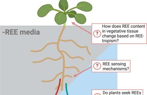
The study, published in Plants, People, Planet, shows how some plant species have evolved to capitalize on the properties of rare earth elements (REEs)—a group of metals essential to the energy transition. Biology has already inspired new metal extraction techniques. For example, “biomining” accounts for approximately 15% of copper mining.

Co-author Professor Caitlin Byrt said we should be looking to biology for inspiration when it comes to efficiently using critical resources, like REEs, to harness and manage clean energy for things like powering our cars and homes.

“Plants are masters of finding efficient strategies for managing and transferring energy. Some plants, for example, exhibit enhanced photosynthesis when they can access REEs,” she said.

“Investigating how plants use these elements in their processes for transferring energy efficiently and precisely could inspire new photon-harvesting devices that mimic photosynthesis, for example.”

REEs are most valuable in their pure forms, which, according to the report’s authors, can be challenging to extract from ores in natural deposits.

“Mechanisms that living organisms use to manage REEs have evolved over millions of years to generate new ways of extracting these valuable resources,” lead author Dr. Samantha McGaughey said.

“Exploration of how REEs interact with biological processes is important for planning the sustainable management of REEs going forward, as we start to rely more and more on these critical resources.

“We need to look at developing innovative approaches to use, reuse and recycle critical resources like REEs and copper to ensure we’re also minimizing negative impacts on the environment. For example, recycling from secondary sources like waste materials can reduce the need for REE extraction from primary sources, using methods that typically lead to disruption of natural ecosystems.”

Professor Byrt added that while there’s exciting potential for using REEs’ resources to manufacture cutting-edge technologies for the future, prioritizing the responsible use of these critical resources is important.

“This means prioritizing the use of critical resources in technologies that improve the quality of life for our communities,” she said.

“Further work in this area of plant science, supported by the Australian Research Council, is expected to help pave the way forward for sustainable use of the resources needed for the clean energy transition.”

Structure of Key Parkinson’s Disease Protein, PINK1, Seen for First Time

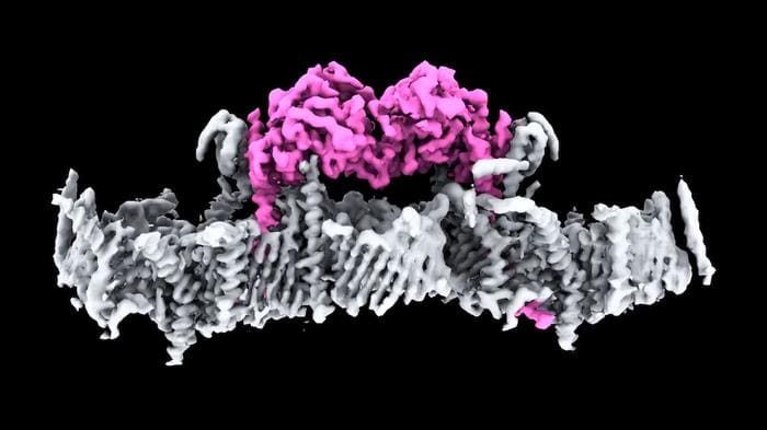
Scientists at Walter and Elizabeth Hall Institute (WEHI) have made what they describe as a huge leap forward in the fight against Parkinson’s disease, solving a decades-long mystery by determining the first structure of the human protein PINK1 bound to mitochondria. First discovered more than 20 years ago, PINK1 is a protein directly linked to Parkinson’s disease, but until now, no one has seen what human PINK1 looks like, or how PINK1 attaches to the surface of damaged mitochondria to be switched on.

David Komander, PhD, head of WEHI’s Ubiquitin Signaling Division, said years of work by his team have now unlocked that mystery. “This is a significant milestone for research into Parkinson’s. It is incredible to finally see PINK1 and understand how it binds to mitochondria,” said Komander, who is also a laboratory head at the WEHI Parkinson’s Disease Research Centre. “Our structure reveals many new ways to change PINK1, essentially switching it on, which will be life-changing for people with Parkinson’s.” Komander is corresponding author of the team’s published paper in Science, titled “Structure of human PINK1 at a mitochondrial TOM-VDAC array.”

Parkinson’s disease is insidious, often taking years, sometimes decades to diagnose. Commonly associated with tremors, there are close to 40 symptoms, including cognitive impairment, speech issues, body temperature regulation, and vision problems. In Australia, more than 200,000 people live with Parkinson’s disease, and between 10% and 20% have a young onset form of the disease (early onset Parkinson’s disease; EOPD), meaning they are diagnosed under the age of fifty years.

One of the hallmarks of Parkinson’s is the death of brain cells. Around 50 million cells die and are replaced in the human body every minute. But unlike other cells in the body, when brain cells die, the rate at which they are replaced is extremely low.

Mitochondria produce energy at a cellular level in all living things, and cells that require a lot of energy can contain hundreds or thousands of mitochondria. The PARK6 gene encodes the PINK1 protein, which supports cell survival by detecting damaged mitochondria and tagging them for removal. “PINK1 is a ubiquitin and Parkin kinase and functions as an early sensor and transducer of mitochondrial damage signaling,” the authors explained.

When mitochondria are damaged, they stop making energy and release toxins into the cell. In a healthy person, when mitochondria are damaged, PINK1 gathers on mitochondrial membranes and signals through a small protein called ubiquitin, that the broken mitochondria need to be removed. In a healthy person, the damaged cells are disposed of in a process called mitophagy. The PINK1 ubiquitin signal is unique to damaged mitochondria, and when PINK1 is mutated in patients, broken mitochondria accumulate in cells.

Study lead author and WEHI senior researcher Sylvie Callegari, PhD, said PINK1 works in four distinct steps, with the first two steps not having been seen before. First, PINK1 senses mitochondrial damage. Then it attaches to damaged mitochondria. Once attached it tags ubiquitin, which then links to a protein called Parkin so that the damaged mitochondria can be recycled. In their paper, the authors explained further, “In healthy mitochondria, PINK1 is translocated across the mitochondrial outer membrane (MOM) via the translocase of the outer membrane (TOM) complex, inserted into the mitochondrial inner membrane (MIM) via the translocase of the inner membrane (TIM)23 complex, cleaved by the MIM protease PARL, retro-translocated and degraded by the proteasome.”

In a person with Parkinson’s and a PINK1 mutation, the mitophagy process no longer functions correctly and toxins accumulate in the cell, eventually killing it. “Mutations in the ubiquitin kinase PINK1 cause early onset Parkinson’s disease,” the authors noted. “A structure of full-length PINK1 at mitochondria is crucial to develop and understand PINK1 activators and treat Parkinson’s disease.”

However, scientists to date have been unable to visualize the protein or understand how it attaches to mitochondria and is switched on. The authors noted that while multiple structures of isolated kinase domains of PINK1 from insects have given scientists molecular details about PINK1 activation, “… human PINK1 has resisted structural characterization, and the PINK1 N terminus which comprises many patient mutations has remained unresolved …”

Commenting on their newly reported work, Callegari said, “This is the first time we’ve seen human PINK1 docked to the surface of damaged mitochondria and it has uncovered a remarkable array of proteins that act as the docking site. We also saw, for the first time, how mutations present in people with Parkinson’s disease affect human PINK1.”

The idea of using PINK1 as a target for potential drug therapies has long been touted but not yet achieved because the structure of PINK1 and how it attaches to damaged mitochondria were unknown. The research team hopes to use the knowledge to find a drug to slow or stop Parkinson’s in people with a PINK1 mutation. “Our structure also provides multiple unexplored avenues to stabilize PINK1 on mitochondria, to develop much-needed treatment options for Parkinson’s disease patients,” the scientists wrote.

Neurons Generated from Skin Cells Without Stem Cell Step in Mice

New research from scientists at the Massachusetts Institute of Technology (MIT) describes a process for converting skin cells directly into neurons that bypasses the induced pluripotent stem cell (IPSC) stage. Details are provided in a pair of papers that were published in Cell Systems. The first paper is titled “Proliferation history and transcription factor levels drive direct conversation to motor neurons.” The second paper is titled “Compact transcription factor cassettes generate functional, engraftable motor neurons by direct conversion.”

Working with mouse cells, the researchers developed an efficient conversion method for producing more than 10 neurons from a single skin cell. According to the study, the researchers successfully generated motor neurons and engrafted them into mouse brains, where they integrated with the host tissue. If they can replicate the process using human skin cells, this could be a way to generate large quantities of motor neurons for cell therapies to treat spinal cord injuries or other neurodegenerative diseases that impair mobility.

“We were able to get to yields where we could ask questions about whether these cells can be viable candidates for the cell replacement therapies, which we hope they could be. That’s where these types of reprogramming technologies can take us,” said Katie Galloway, PhD, senior author on both studies and a professor in biomedical engineering and chemical engineering at MIT.

The established way of reprogramming cells is to use four transcription factors that coax them to become IPSCs. These can then be differentiated into other cell types of interest. The process takes several weeks and many cells fail to fully transition to mature cell types, remaining stuck in intermediate states. To avoid this, Galloway and her colleagues came up with a way to directly convert somatic cells to neurons without the intermediate iPSC step.

In a previous study, the scientists demonstrated this type of direct conversion, but with very low yields—less than 1%. That approach used a combination of six transcription factors plus two other proteins that stimulate cell proliferation. Each of the eight genes that encode for these projects was delivered using a separate viral vector. And that made it difficult to ensure that each was expressed at the correct level in each cell.

Galloway and her team have now streamlined the process so that skin cells can be converted to motor neurons using just three transcription factors, plus the two genes. Using mouse cells, the researchers experimented with different combinations of the original six transcription factors. They dropped one factor at a time until they reached a combination of three—NGN2, ISL1, and LHX3—that successfully accomplished the conversion from skin cells to neurons.

Once they narrowed the number of relevant genes down to three, the researchers used a single modified virus to deliver all three, ensuring that each cell expresses each gene at the correct levels. Using a separate virus, the researchers also delivered genes that encoded for p53DD and a mutated version of HRAS. These genes drive the skin cells to divide many times before they start converting to neurons, allowing for a much higher yield of neurons.

That last step is important because “if you were to express the transcription factors at really high levels in non-proliferative cells, the reprogramming rates would be really low, but hyperproliferative cells are more receptive. It’s like they’ve been potentiated for conversion, and then they become much more receptive to the levels of the transcription factors,” Galloway explained.

The researchers have now developed a slightly different combination of transcription factors that allows them to perform this direct conversion in human cells but with a lower efficiency rate—between 10–30%. This process takes about five weeks, which is slightly faster than converting the cells to iPSCs first and then turning them into neurons.

Once they identified the optimal combination of genes to deliver, the researchers also worked on improving their delivery mechanism. After testing three different delivery viruses, they found that a retrovirus achieved the most efficient rate of conversion. Reducing the density of cells grown in the dish also helped to improve the overall yield of motor neurons.

As part of their study, the researchers tested whether the motor neurons generated by their process could be engrafted into mice brains. They delivered the cells to the striatum, a part of the brain involved in motor control and other functions. Two weeks later, many of the neurons had survived and seemed to be forming connections with other brain cells. The cells also showed measurable electrical activity and calcium signaling, which suggests that they may be able to communicate with other neurons.

The researchers now plan to explore the possibility of implanting these neurons into the spinal cord. They also hope to increase the efficiency of this process for human cell conversion.

error: Content is protected !!