Perimenopause explained: What it is, plus products that may help with symptoms

When it comes to health trends, menopause is having a long-overdue moment — and for good reason. It’s something half the population will experience, yet it’s been historically under-discussed. In fact, one study found that only 31% of doctors received menopause training in medical school.

Because of that, many women aren’t familiar with the stage that comes before menopause: perimenopause.

Perimenopause is when hormone levels — especially estrogen and progesterone — begin to fluctuate in the years leading up to menopause. And with that shift can come a host of confusing, sometimes disruptive symptoms, from hot flashes and night sweats to skin changes and mood swings.

Below, doctors explain what perimenopause is, how to tell if you’re in it, and what you can do to ease the transition. Plus, we share expert-recommended products that may help with common symptoms.

What is perimenopause?

Perimenopause refers to the transitional phase before menopause, according to the National Library of Medicine. It’s marked by shifting reproductive hormone levels — especially estrogen and progesterone — and typically starts in a woman’s 30s to 50s.

There’s no single test that confirms perimenopause, since hormone levels naturally fluctuate throughout the month. Instead, doctors assess your age, symptoms, and family history to make a diagnosis.

Symptoms of perimenopause

Perimenopause looks different for everyone — some experience intense symptoms, while others have a more gradual transition. Research suggests it begins about 5 to 10 years before menopause. Common symptoms include:

  • Hot flashes and night sweats: Caused by hormonal shifts affecting the hypothalamus, your body’s temperature regulator.
  • Irregular periods, vaginal dryness, and low libido: Estrogen and progesterone imbalances can impact the menstrual cycle and sexual health.
  • Skin and hair changes: Lower estrogen levels reduce collagen and moisture, leading to dryness, thinning skin, and increased hair shedding.
  • Mood swings and brain fog: Hormonal fluctuations can mimic the emotional volatility of adolescence.
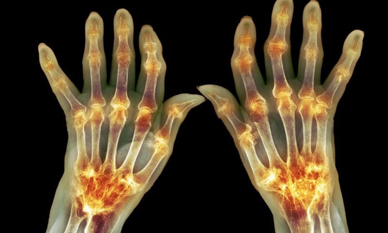
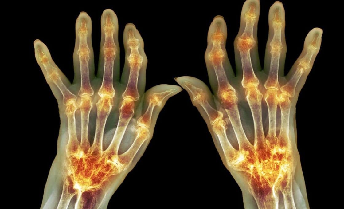
  • Sleep issues, weight gain, joint pain, and frequent urination are also commonly reported.

What can you do about perimenopause symptoms?

You don’t have to tough it out — there are actionable ways to ease symptoms.

“The best thing you can do is approach aging with intention,” says Dr. Cristina Del Toro Badessa, a board-certified physician in integrative medicine. That includes eating well, exercising, limiting alcohol and caffeine, managing stress, and prioritizing sleep and relationships.

Some women benefit from supplements like collagen peptides or biotin, which may support skin and hair health. Others explore hormone replacement therapy (HRT), though it’s best discussed with a doctor.

There are also targeted products that can provide comfort and relief for specific symptoms — a few of our expert-approved picks are below.

Our top picks for perimenopause symptoms

While over-the-counter products are unlikely to eradicate symptoms, several things can make them feel more manageable. The following items were all created to address common perimenopause symptoms and are either highly recommended or recommended by an expert.

NBC Select reporter Zoe Malin is a fan of this personal cooling device and it can be a great tool to help you combat sudden hot flashes. The rechargeable fan is meant to be worn around your neck and, when turned on, it has vents that blow cool air toward you. It is also bladeless, so you don’t need to worry about hair getting caught. This fan has three airflow speeds, and you can change the angle of the vents to help the air blow where you want it.

Dealing with night sweats? These cooling sheets made our list of the best bed sheets. They are made with a cool-to-the-touch, moisture-wicking fabric to help regulate body temperature and mitigate sweat, according to the brand. Also lovely: They get softer the more you wash them, according to Sheex.

Experts previously told us it’s important to prioritize skin care products with hydrating ingredients when in perimenopause. This serum, which is made specifically for women in that stage of life, contains fatty acids to moisturize and vitamin C to brighten. It should be applied in the morning before using SPF and moisturizer.

If you’ve noticed perimenopause has led to dry, brittle hair, switching to a super hydrating conditioner can help. This one contains hydrating cupuaçu butter, which can also tame frizz and add shine, according to Biolage.

Ease joint aches with a massage gun. This one won an NBC Select Wellness Award last year and is a favorite amongst Select editors, including editorial director Lauren Swanson, who tested massage guns while marathon training. She said this one had heads and speeds that were gentle on tender muscles. It also has an ergonomic handle, making it easier to hold as you run it over your body and an LCD screen that makes the settings easy to read.

Children’s Cancer Risk From Gas Stoves Nearly Double That of Adults

The gas stove in your home could be releasing toxic chemicals at cancer-causing levels, a new study shows. The study focuses on benzene, which is produced from the burning of natural gas or propane, and is already known to increase cancer risk.

Researchers led by a team from Stanford University measured benzene emission rates from gas stoves in 87 homes, as well as analyzing the movements of the gas in a smaller number of locations, and running computer models of how benzene might spread.

For homes with stoves that emit the most benzene and have the least ventilation, benzene-caused cancer risks are “significantly elevated”, the data showed – especially for children, where the lifetime risk rose 1.85 times higher than it did for adults.

“Natural gas and propane stoves emit benzene, a known carcinogen through combustion,” write the researchers in their published paper. “This study evaluates population-level benzene exposure and associated health risks for the 6.3 million U.S. residents exposed to the top 5 percent highest benzene-emitting gas stoves.”

The study produced numerous data points. Cancer risks were greater for apartments and smaller homes, as you might expect, while benzene often spread far enough to reach bedrooms – further increasing the danger, as that’s where we spend much of our time.

As per the World Health Organization (WHO), an acceptable cancer risk for exposure is seen as one in a million: so if a million people are given that level of exposure, a single person might be expected to develop cancer because of it.

This analysis showed that for the bedroom exposure from the worst benzene-emitting gas stoves, the lifetime cancer risk was 1.92-12.03 in a million for kids, and 0.94-5.89 in a million for adults – far over WHO’s recommended levels.

It’s important to note that these are worst-case scenarios – the highest benzene emissions and the lowest levels of ventilation – but they highlight the potential dangers, especially for children, who have lower body weights and faster breathing rates.

“These values may vary depending on home size, weather circumstances, the usage of a hood, ventilation in the home, and other housing factors, the frequency and duration of using ovens and burners, and vary from person to person metabolic and other physiological characteristics,” write the researchers.

That is the good news from this study. Increasing ventilation (even just opening a window) and reducing how often gas stoves are used makes a big difference, and that’s something the researchers are keen to raise awareness of.

The researchers also put their findings into some useful context: in the US, people spend an average of 90 percent of their time indoors, and with that figure rising with more of us working from home, further indoor air pollution studies are needed.

“The study underscores the importance of addressing combustion-related indoor air pollutants to protect public health, particularly in households with limited ventilation,” write the researchers.

The research has been published in the Journal of Hazardous Materials.

Beyond T Cells: Harnessing Innate Immunity to Prevent Transplant Rejection

When a transplanted organ arrives, it’s like a controlled burn that risks becoming a wildfire. The body’s innate immune system senses damage signals, like heat shock proteins (HSP70), and sounds the alarm, mobilizing dendritic cells to fan the flames of inflammation. Antigen-presenting cells rally T cells to the scene, launching an attack on the foreign tissue. These are the first steps of organ transplant rejection.

Current immunosuppressants act like firefighters putting out the flames once the building (in our case, the tissue) is a blaze. In a new study, Siglec-E in mice (and Siglec-7/9 in humans) acts as an early intervention alert, preventing those fires from spreading in the first place. This receptor binds sialic acid ligands to keep dendritic cells from overreacting. By blocking NF-κB signaling and dampening pro-inflammatory cytokines like TNF-α, Siglec-E keeps the immune response from spiraling out of control.

Without these inhibitory receptors, the immune response surges unchecked, leading to heightened inflammation, accelerated T-cell activation, and faster transplant rejection. By targeting this upstream checkpoint, the researchers propose, it may be possible to quiet the inflammatory blaze at its source—offering a possible therapeutic strategy to protect transplanted organs without broadly suppressing immune function.

Innate immunity’s role in rejection

Current immunosuppressive therapies primarily target T cells, the drivers of the adaptive immune response that recognizes and attacks transplanted organs. These treatments, while effective at reducing rejection, come with a high cost: they broadly suppress immunity, leaving patients vulnerable to infections, cancers, and other complications. Yet despite this aggressive suppression of T cells, many transplants still fail over time. Increasing evidence suggests that early inflammation, mediated by the innate immune system, plays a pivotal role in setting the stage for rejection.

Recognizing this gap, the Mass General Brigham researchers turned their attention upstream, to the body’s first line of defense. Rather than focusing solely on dampening T cells, they explored whether controlling the innate immune response could prevent the inflammatory cascade from spiraling in the first place. By investigating the inhibitory receptor Siglec-E in mice—and its human counterparts, Siglec-7 and Siglec-9—they identified a natural checkpoint that calms overactive immune responses early on.

To test the role of Siglec-E, the team used preclinical mouse models of heart, kidney, and skin transplantation. They found that mice lacking Siglec-E experienced accelerated rejection, heightened inflammation, and increased activation of dendritic cells—the antigen-presenting cells that bridge innate and adaptive immunity. Without Siglec-E, dendritic cells stayed hyperactivated, producing more pro-inflammatory cytokines like TNF-α and IL-6, and driving stronger T-cell responses against the allograft.

When the researchers analyzed human transplant samples, they observed that higher levels of Siglec-7 and Siglec-9 were associated with better graft survival. This finding suggests that the protective role of these inhibitory receptors extends to humans, offering translational potential for new therapies.

By identifying this natural “brake” in the immune system, the study points to a new therapeutic strategy: targeting Siglec-7 and Siglec-9 to modulate dendritic cell activation, reducing inflammation without globally shutting down the immune system. Instead of waiting to suppress T cells after the immune system is already fully mobilized, therapies directed at this pathway could quiet the alarm at its source, preventing the inflammatory cascade that leads to rejection.

A promising target for next-generation transplant therapies

“Regulating innate immune activation is a crucial step in the prevention of transplant rejection and improvement in transplant outcomes,” the researchers state. Prolonged innate immune responses may reduce tolerance while interfering with trained immunity and the adaptive immune response. To maintain a balanced immune response, the innate immune response must be recognized as a front-line defender against excessive inflammation and possible tissue damage.

“By harnessing natural inhibitory pathways like Siglec-E, we can develop safer, more precise therapies that protect transplanted organs without compromising overall immune health,” said Leonardo Riella, MD, PhD, medical director of kidney transplantation at Massachusetts General Hospital.

As a negative regulator of innate immune responses and acute T cell-mediated transplant rejection in mice, Siglec-E offers a potential therapeutic target that may be translatable to humans. This offers hope for a next generation of organ transplant treatments, including longer-lasting transplant success and reducing the need for lifelong immunosuppression.

In Vivo Bioprinting Shows Promise for 3D Printed Implants Without Surgery

Researchers headed by a team at the California Institute of Technology developed an ultrasound-guided 3D printing technique that could make it possible to fabricate medical implants in vivo and deliver tailored therapies to tissues deep inside the body—all without invasive surgery. The researchers say the imaging-guided deep tissue in vivo sound printing (DISP) platform utilizes low-temperature–sensitive liposomes (LTSLs) as carriers for cross-linking agents, enabling precise, controlled in situ fabrication of biomaterials within deep tissues.

Reporting on their development in Science (“Imaging-guided deep tissue in vivo sound printing”), first author Elham Davoodi, PhD, and senior, corresponding author Wei Gao, PhD, described proof of concept studies demonstrating in vivo printing within the bladders and muscles of mice, and rabbits, respectively. Gas vesicle (GV)–based ultrasound imaging integrated into the printing platform enabled real-time monitoring of the printing process and precise positioning. In their paper, the authors concluded, “DISP’s ability to print conductive, drug-loaded, cell-laden, and bioadhesive biomaterials demonstrates its versatility for diverse biomedical applications.”

Three-dimensional (3D) bioprinting technologies offer significant promise to modern medicine by enabling the creation of customized implants, intricate medical devices, and engineered tissues, tailored to individual patients, the authors wrote. “However, the implantation of these constructs often requires invasive surgeries, limiting their utility for minimally invasive treatments.”

While in vivo bioprinting—“3D printing” tissue directly within the body—offers a less invasive alternative, it has been limited by challenges such as poor tissue penetration depth, a narrow range of biocompatible bioinks, and the need for printing systems that operate at high resolution with precise, real-time control. “Although near-infrared (NIR) light has been explored as a biosafe energy source for in vivo printing, its applications remain restricted to subcutaneous tissues due to limited light penetration,” the team continued.

To address these barriers, Davoodi and colleagues developed a novel imaging-guided platform, imaging-guided DISP, which uses focused ultrasound and ultrasound-responsive bioinks to precisely fabricate biomaterials directly within the body. These bioinks, or US-inks, combine biopolymers, imaging contrast agents, and temperature-sensitive liposomes carrying crosslinking agents and can be delivered to targeted tissue sites deep within the body via injection or catheter. “US-inks are composed of biopolymers, cross-linking agent-encapsulated LTSLs, and GVs that act as ultrasound imaging contrast agents,” the team further explained. “These bioinks are delivered to the target sites through injection or catheters and are located using an ultrasound imaging setup integrated into a 3D printing platform.”

A focused ultrasound (FUS) transducer, guided by automated positioning and a predefined digital blueprint, triggers localized low-temperature heating (slightly above body temperature) that releases the crosslinker, initiating immediate in situ gel formation. “Localized heating induced by FUS triggers the release of cross-linking agent from the LTSLs, enabling immediate in situ cross-linking of the US-ink.” The bioinks and their resulting gels can be tailored for various functions, including conductivity, localized drug delivery, and tissue adhesion, as well as real-time imaging capabilities.

Davoodi and colleagues validated the DISP technology by successfully printing drug-loaded and functional biomaterials near cancerous sites in a mouse bladder and also deep within rabbit muscle tissue, demonstrating potential applications for drug delivery, tissue regeneration, and bioelectronics. “We validated DISP by successfully printing near diseased areas in the mouse bladder and deep within rabbit leg muscles in vivo, demonstrating its potential for localized drug delivery and tissue replacement,” the team stated.

Further biocompatibility tests revealed no signs of tissue damage or inflammation, and the body cleared unpolymerized US-ink within a week, illustrating the platform’s safety. “The DISP technology offers a versatile platform for printing a wide range of functional biomaterials, unlocking applications in bioelectronics, drug delivery, tissue engineering, wound sealing, and beyond,” the team stated. “By enabling precise control over material properties and spatial resolution, DISP is ideal for creating functional structures and patterns directly within living tissues.”

In a related perspective, Xiao Kuang, PhD, at the University of Wisconsin-Madison, wrote, “Although Davoodi et al. advanced ultrasound 3D printing toward clinical translation, additional refinements are needed to implement the technology for clinical use … A detailed relationship between process conditions, the structure of the printed material, and the resulting properties must be elucidated through careful testing.”

Novavax Sales Jump 600% YoY as COVID-19 Vaccine Still in Limbo at FDA

After more than a month of speculation over how the FDA will rule on Novavax’s application for an updated formulation of its COVID-19 shot, CEO John Jacobs said on the company’s Q1 earnings call: “When we have it, we’ll have it. Until then, we don’t have it.”

Novavax’s updated COVID-19 vaccine has been in limbo after the FDA missed its PDUFA deadline and subsequently requested another trial—though it’s unclear if that trial needs to happen before approval. But today, the company gave investors some good news, reporting a massive year-over-year increase in sales for the original product on a first-quarter earnings call Thursday.

In its Q1 2025 earnings report, the Maryland-based company reported $667 million for the quarter, an increase of 610% over the $94 million brought in during the first three months of last year. The majority of this revenue came from sales of the COVID-19 vaccine, Nuvaxovid, which clocked $608 million—a dramatic increase over the $82 million it generated in Q1 2024.

Nuvaxovid was the primary theme running through the call, in which executives also touched on the company’s earlier-stage pipeline—including a combination COVID-19/flu vaccine currently in Phase III studies—as well as partnerships and a recently reported reactogenicity study.

On April 1, the FDA did not announce a decision regarding full approval of Nuvaxovid, which is currently marketed under an emergency use authorization (EUA). The missed PDUFA ignited significant controversy and placed Novavax at the heart of the vaccines debate currently broiling under Health and Human Services Secretary Robert F. Kennedy Jr.

At one point during the Q&A period, which was riddled with questions regarding the vaccine’s BLA, CEO John Jacobs appeared to show some exasperation, saying: “When we have it, we’ll have it. Until then, we don’t have it.”

On April 25, The Wall Street Journal reported that the FDA was requesting the company complete an additional randomized clinical trial, a fact Novavax has confirmed. While executives have maintained—including on Thursday’s call—that this would be a postmarketing study, FDA Commissioner Marty Makary raised questions when he posted on X: “To be clear, this is a new product that Novavax is trying to introduce to the market with a study of a different product from 2021. New products require new clinical studies.”

Makary’s statement presaged a new policy revealed by HHS last week requiring all new vaccines to be tested in placebo-controlled trials before they are approved.

Jacobs sought to clarify any ambiguity in responding to a question regarding the potential for an additional clinical trial prior to approval of the BLA. “Based on what we’ve received to date, formally from FDA, they’re asking for a postmarketing commitment, and by definition, it’s our understanding that a postmarketing commitment comes after approval when you’ve begun to market that product,” he said.

Novavax CFO Jim Kelly added that the company is “working closely with our partner Sanofi to determine the potential approach to funding” the postmarketing study. Notably, Novavax did not provide full-year 2025 guidance for sales of Nuvaxovid, and Kelly said that potential costs of a postmarketing study should be considered in terms of Novavax’s multi-year guidance.

Despite the recent attention on the COVID-19 vaccine, Jacobs said that Novavax has now pivoted focus to its partnerships, including the licensing agreement with Sanofi, which Jacobs called a first priority. Signed in May 2024, the deal covers the co-commercialization of Nuvaxovid and development of the novel COVID-19/flu combo shot.

Meanwhile, Novavax touted the results of the SHIELD-Utah study, comparing the updated JN.1 Nuvaxovid shot to Pfizer and BioNTech’s Comirnaty in terms of reactogenicity. Preliminary data, reported on April 15, showed that Novavax’s protein-based vaccine “induced lower frequency and severity of short-term side effects and impact on daily life” compared to the mRNA-based Comirnaty.

Novavax raised its full-year 2025 guidance from $975 million to $1.03 billion.

Vor Bio Looks for Exit in Challenging Funding Environment; Lays Off 95% of Staff

The cell engineering company, co-founded by oncologist and writer Siddhartha Mukherjee, does not see a path forward for its pipeline of early-stage cell therapies for two different types of cancer.
Cambridge, Massachusetts–based Vor Bio is bowing out amid a tough funding environment after examining the clinical data currently available for its assets. The company, co-founded in 2015 by oncologist and Pulitzer Prize-winning author Siddhartha Mukherjee based on work from his Columbia University laboratory, was focused on cell therapies for acute myeloid leukemia and myelodysplastic syndrome.

The company will now cease all clinical and manufacturing activity, including ongoing clinical trials, and look for ways to “maximize shareholder value.” This could include licensing or selling its assets, executing a merger, selling the company outright or some other “strategic action,” according to a Thursday announcement. The decision to wind down operations is based entirely on the company’s available clinical data and a “challenging fundraising environment,” according to the release.

With the announcement, Vor is also letting go of 95% of its staff, incurring $10.9 million in related costs. According to a recent SEC filing, Vor had 159 full-time employees as of March 2025. The company will retain eight employees for the time being to keep the lights on while they are “maintaining compliance with regulatory and financial reporting requirements, and winding-down the clinical and manufacturing operations.”

Vor had $91.9 million in cash, cash equivalents and marketable securities on hand as of the end of 2024, according to Vor’s statement. There is no timeline for this strategic shift and the company “does not intend to discuss or disclose further developments during this process” until a specific action is approved by the board of directors.

As recently as December 2024, Vor raised $55.6 million through a PIPE financing arrangement led by LinkedIn co-founder Reid Hoffman. That money was to go toward advancing Vor’s pipeline of cell therapy oncology treatments for acute myeloid leukemia (AML) and myelodysplastic syndrome (MDS). Those treatments included an engineered cell transplant called Trem-cel for AML and MDS as well as a CAR T treatment called VCAR33 for AML.

Both of those therapies were in Phase I/II trials, according to Vor’s published pipeline. Vor also has antibody-drug conjugates and cell transplants for treating AML, which were in preclinical discovery and IND-enabling studies.

AI Uncovers Gut Signature Tied to Complex Regional Pain Syndrome

By training machine learning algorithms on data from gut bacteria, scientists from McGill University and their collaborators developed a computational tool to detect patterns in the microbiome that are connected to complex regional pain syndrome (CRPS), a relatively rare condition that affects hundreds of thousands of people worldwide. When they applied the tool to patient samples, it identified a common microbiome signature connected to CRPS n that could lead to advances in how the condition is treated. Details are provided in a new Anesthesiology paper titled, “Altered gut microbiome composition and function in individuals with complex regional pain syndrome.”

CRPS typically develops in a limb after injury or surgery and can lead to long-term disability. It causes severe, persistent pain that is often far worse than the initial injury, along with swelling and changes in skin color and temperature. The condition is challenging to treat, “with patients often experiencing prolonged suffering before receiving appropriate care,” said Amir Minerbi, MD, PhD, senior author on the paper, director of the Institute for Pain Medicine in Haifa, Israel, and senior lecturer at the Technion–Israel Institute of Technology.

The results of this study could help change that. As the researchers noted in the paper, even though the condition has been linked to “dysregulation in several physiologic systems … including aberrant inflammatory and immune responses, vasomotor dysfunction, and nervous system changes,” the exact cause remains unclear, making it harder to treat. Their research could “pave the way for future studies elucidating the pathophysiology of CRPS and exploring new diagnostic aids and treatment modalities.”

Digging into the details, the scientists explain that they used advanced machine learning to analyze gut microbiome samples from two cohorts from Israel and Canada—the samples from Israel were used for training. In total, the scientists analyzed data from 120 microbiome samples as well as over 100 plasma samples from 53 CRPS patients and 52 unrelated controls.

The scientists reported that their algorithm identified significant differences between the gut bacteria of CRPS patients and pain-free individuals. In fact, they successfully predicted CRPS in Canadian patients with over 90% accuracy, according to Emmanuel Gonzalez, PhD, lead author on the paper and a member of the McGill Centre for Microbiome Research. “This is extraordinary because factors like geography, climate, diet, and natural variation between people typically create large microbiome differences. Yet, our AI approach seems to have identified a common ‘microbiome signature’ of CRPS, suggesting microbiome-based diagnostics could work across populations in different countries.”

Furthermore, even patients whose symptoms cleared after limb amputation still had the same gut bacteria pattern linked to CRPS. That suggests that the gut microbiome “might make some people more prone to developing CRPS, with an injury or other event triggering the condition,” said Yoram Shir, MD, a professor in the department of anesthesia at McGill’s Faculty of Medicine and Health Sciences.

Microscopy method can reconstruct mammalian brain tissue in synaptic detail

Our brain is a complex organ. Billions of nerve cells are wired in an intricate network, constantly processing signals, enabling us to recall memories or to move our bodies.

Making sense of this complicated network requires a precise look into how these nerve cells are arranged and connected. “LICONN,” a new microscopy method developed by scientists at the Institute of Science and Technology Austria (ISTA) and Google Research, now helps piece together this puzzle.

Light microscopes have been evolving for centuries. Scientists use light microscopy to—literally and figuratively—illuminate the most intricate biological structures. However, unraveling the complex details and architecture of the brain remains a seemingly impossible challenge, considering its billions of densely packed neurons, each linked to other cells via thousands of synapses.

A new microscopy pipeline called “LICONN” (light-microscopy-based connectomics), developed at the Institute of Science and Technology Austria (ISTA), now offers a breakthrough.

LICONN is the first technology beyond electron microscopy capable of reconstructing brain tissue with all the synaptic connections between neurons. It also opens up the possibility of visualizing complex molecular machinery alongside the structure of neurons, all while utilizing standard light microscopes for measurements.

This new technique was developed by Mojtaba R. Tavakoli, Julia Lyudchik, Johann Danzl, and their colleagues from the High-Resolution Optical Imaging for Biology research group at ISTA. They collaborated with the Novarino group at ISTA and Michal Januszewski and Viren Jain from Google Research.

The method is now published in Nature.

New possibilities with LICONN

Mojtaba R. Tavakoli opens a curtain, revealing a light microscope with endless wires connecting the optical instrument to a computer. The screen’s lights shine bright—glooming shades of green and pink illuminate the almost pitch-black room.

“That’s the hippocampus—a brain region responsible for memory formation,” says Tavakoli and points to the screen. “The fluorescent dots you see are molecules involved in synaptic transmission.” The ISTA graduate moves the frame and adjusts the settings.

LICONN is the Danzl group’s newest microscopy technique. It acts like a meticulous puzzle solver, assembling the intricate brain networks by piecing together the finest neuronal processes and correctly linking each synaptic connection to its respective neuron.

“Up to now, no light microscopy technique could do that,” says Johann Danzl, a trained medical doctor and physicist, now professor at ISTA. “It was a longstanding goal of our group to build such a pipeline for reconstructing brain tissue. And LICONN can do this while placing specific molecules into the context of the structural reconstruction.”

What stands out is that the image acquisition is done on a standard off-the-shelf microscope, which is very fast and offers multicolor capability.

The technique can be reproduced anywhere in the world, as scientists do not require high-end, expensive equipment that would be needed for current approaches for brain tissue reconstruction. To obtain this level of detail, the resolution has to be extraordinarily high, around a few tens of nanometers, 10,000 times smaller than the width of a human hair. But how to accomplish that? Expertise in chemistry comes in handy.

Zooming with a gel

For LICONN, the team made use of the chemical and physical properties of hydrogel, a three-dimensional polymer network. Hydrogel has similar characteristics to baby diapers: it can take up water and swell, but does so in a highly controlled manner.

The brain tissue of interest is embedded in this hydrogel. “Cellular components are linked to the hydrogel, meaning the cells’ fine ultrastructure is imprinted onto the gel and preserved for microscopy,” explains Danzl. Before imaging, the structures are expanded by adding water to the material.

As a result, the gel elongates in size in every direction but maintains the relative spatial arrangements of the tissue’s structures with extremely high fidelity.

For comparison, traditional light microscopes are classically limited in their resolving power to around 250–300 nanometers. While this is adequate to visualize larger cellular structures, it is insufficient to reconstruct the densely packed brain tissue.

“The hydrogel expansion pulls features of the brain tissue so far apart that we can resolve them with a standard light microscope. This method enhances the effective resolution by 16 times, achieving a resolution better than 20 nm,” Tavakoli explains.

Research at the intersection of disciplines

Neuroscience and chemistry were not the only fields that found their way into this project. Methods from computer science played a crucial part in the pipeline’s development. This is because capturing microscopic images results in the collection of numerous data points. As such, the intricacy of the datasets reflects the brain’s complexity.

Thus, manually interpreting and reconstructing all the neuronal structures on a sizable scale would be far too laborious. Therefore, Google Research’s deep-learning techniques were trained to segment the individual cells in the tissue.

“Automating the identification of neurons and their elaborate structures on a wider scale using artificial intelligence made the daunting task of reconstructing all the cellular components practically tractable,” explains Viren Jain from Google Research. “The ability to concomitantly visualize specific molecules adds a new quality of information.”

Julia Lyudchik, a Ph.D. student and computer scientist in the Danzl group, played an instrumental role in interpreting the complex datasets.

“Thanks to the exceptionally high resolution of the data, it was possible to automatically detect the synaptic connections between neurons and to transform raw brain imaging data into detailed connectivity maps. This is a complex image processing challenge,” Lyudchik explains.

“In addition, the methods had to be both efficient and scalable, given that even a small piece of brain tissue can contain tens of thousands of synaptic connections.”

LICONN makes it possible to map the location of specific molecules onto neuronal reconstructions, such as those involved in the transmission of signals between neurons at synapses. Lyudchik’s artistic vein helped her create stunning 3D renderings of the brain network, as visualizations are powerful tools to make complex scientific data more accessible and interpretable.

Unlocking new details in the brain’s architecture

By following this comprehensive pipeline, scientists can meticulously reconstruct brain tissue and visualize neuronal connections and networks.

The interplay between experimentation and analysis across disciplines—from imaging and experimentation at ISTA to Google Research’s application of advanced deep learning technologies and the computational analysis at ISTA—results in 3D visualizations of the brain’s architecture at a new level of complexity.

“LICONN brings us a step closer to assembling the puzzle pieces of the mammalian brain and better understanding its functioning both in health and disease,” Danzl concludes.

New screening method finds novel approaches to combat antimicrobial resistant bacteria

Scientists from the Ineos Oxford Institute for antimicrobial research (IOI) have developed a new screening method to tackle bacterial resistance to the tetracycline class of antibiotics. The results from this method provide the starting point for developing new drugs to treat drug-resistant infections. The findings have been published in Chemical Science.

Tetracyclines are among the most widely used antibiotics to treat respiratory tract infections, sexually transmitted diseases, and urinary tract infections. Tetracyclines are also used as growth promoters in livestock production. However, bacteria are becoming resistant to tetracycline antibiotics by producing an enzyme called Tet(X). This enzyme breaks down tetracycline antibiotics, rendering them ineffective against bacteria.

One effective strategy to restore the activity of the antibiotic is to use a combination therapy to counter bacterial resistance mechanisms. An antibiotic combination treatment includes an antibiotic and an inhibitor. The inhibitor prevents bacterial enzymes such as Tet(X) from breaking down the antibiotic before it has its desired effect to treat the infection.

Scientists at the IOI have developed a fluorescent tetracycline probe that can bind to Tet(X). When the probe binds to the enzyme, a change in the fluorescent light emitted by the probe can be measured. Molecules that act as an inhibitor for Tet(X) cause the probe to become displaced from the enzyme, which alters the fluorescent signal.

By measuring changes in fluorescence, researchers can screen high volumes of compounds to quickly and reliably identify those that show promising inhibitory activity against Tet(X).

Using this new experiment, the team screened thousands of existing drugs and identified six promising Tet(X) inhibitors, including molecules already used as antipsychotics, antimalarials, and gut motility drugs.

The antipsychotic trifluoperazine, its chemical cousin prochlorperazine, and the serotonin receptor agonist tegaserod were found to bind inside Tet(X)’s active site by X-ray crystallography. The crystallography allowed the researchers to look at the three-dimensional structure of Tet(X) with the inhibitors bound to understand how they block the enzyme from destroying tetracyclines. This can provide a basis from which to design new inhibitors.

Professor Christopher Schofield, Director of Chemistry, Ineos Oxford Institute for antimicrobial research and senior author of the paper said, “The global rise of Tet(X)-mediated resistance threatens to undermine the effectiveness of last-line antibiotics. Pairing these antibiotics with inhibitors that block enzyme degradation is essential to protect these drugs.

“This is a strategy that has been extremely successful with the penicillin antibiotics, for example augmentin, but has not been adopted on other classes of antibiotics. We have found promising compounds and developed a robust assay platform to accelerate development of tetracycline inhibitors—laying the groundwork for next-generation combination therapies.”

Dr. Matthew Beech, Postdoctoral Research Associate, Ineos Oxford Institute for antimicrobial research and first author of the paper said, “Our newly-developed fluorescent probe has helped us discover existing medicines such as antipsychotics and antimalarials that can be used to protect tetracycline antibiotics.

“Crystal structures have also revealed how these compounds latch onto Tet(X), unlocking new design strategies. We will now work to refine these molecules, with the ultimate aim of delivering a new combination therapy that can be used in clinical settings.”

Gut Microbiome Linked to Rheumatoid Arthritis Through Reprogrammed T Helper Cells

After spending years tracing the origin and migration pattern of an unusual type of immune cell in mice, researchers headed by a team at The Ohio State University College of Medicine have shown how the activity of “good” microbes in the gut is linked to rheumatoid arthritis (RA) and, potentially, other autoimmune diseases.

Scientists first reported in 2016 that specific gut microbes known as commensal bacteria, which cause no harm and often contribute to host health, set off production and release of a gut-originated T cell that drives up body-wide autoimmune disease in mice. Since then, the team has focused on explaining this unexpected twist in the typically harmonious relationship between these microbes and the body.

The gut is where the action begins, but the overall outcome can be attributed to T cells’ “plasticity”—their flexibility to respond to a changing environment, such as in our body’s barrier, the gut.

The research indicates that reprogrammed T helper cells adopt characteristics of a new T helper cell type while preserving some of their original traits, making them “super powerful and potent—and if you are dealing with autoimmune disease, that’s bad news,” said senior and corresponding study author Hsin-Jung Joyce Wu, BVM, PhD professor of internal medicine, division of rheumatology and immunology, at The Ohio State University College of Medicine. “This is really the first time it’s been shown that T cell plasticity, which typically occurs in the gut, can have this dramatic impact outside the gut with systemic impact on autoimmune disease.”

The newly reported findings likely have relevance to human patients, Wu said. Many of the gene expressions detected in these abnormal cells in mice also exist in the same cells in people with rheumatoid arthritis. Wu is senior author of the team’s paper in Nature Immunology, titled “Aberrant T follicular helper cells generated by TH17 cell plasticity in the gut promote extraintestinal autoimmunity.” In their paper the team concluded, “Our findings offered a mechanism whereby T cell plasticity, a process originating in the gut and aided by gut microbiota, powerfully promotes autoimmunity at gut-distal sites.”

“Autoimmune diseases have risen steeply in the industrialized world,” the authors wrote. An estimated 18 million people worldwide are affected by rheumatoid arthritis, a chronic autoimmune disease-causing inflammation throughout the body and pain in the joints. Like other autoimmune diseases, RA is caused by the immune system attacking the body’s tissues and organs. Though the exact cause is unknown, genetics and environmental exposures—such as smoking and changes of gut commensal bacteria, or dysbiosis—are among the risk factors.

The abnormal T cells in question is called a T follicular helper 17 (TFH17) cell—meaning it functions as a TFH cell but also displays T helper 17 (TH17) cell signatures. Several previous studies have reported that the human equivalent of these types of cells are found in the blood of patients with autoimmune diseases, and are linked to more severe symptoms, but little has been known about the cells’ backstory. “An excessive TFH cell response can lead to overproduction of autoantibodies (auto-Ab) and autoimmunity,” the team further noted, while “Much remains unknown regarding T follicular helper 17 (TFH17) cells commonly found in autoimmune patients.”

These cells have been a puzzle, Wu said, because the conventional TFH cells are expected to be nonmobile, and just reside in B cell follicles to help B cells, another immune cell type critical for the development of RA. “T follicular helper (TFH) cells are a subset of CD4+ T cells, specializing in B cell help,” they pointed out. “Unlike other T effector cells, conventional TFH cells are not very mobile as they are mostly confined to the B cell follicles where they originated.”

However, in contrast with conventional TFH cells, the TFH17 cells also have the traveling capabilities of T helper 17 cells, which are known to migrate rapidly to infection sites where they produce the proinflammatory protein IL-17. “Circulating T follicular helper 17 (cTFH17) cells are a subset of T cells sharing both TFH and T helper 17 (TH17) cell signatures, which are found in the blood of numerous types of autoimmune patients,” the investigators explained.

Following their 2016 study, work in Wu’s lab has now discovered that the systemic TFH cells traced back to Peyer’s patches (PP), lymphoid tissue in the small intestine, and induced by typically harmless microbes called segmented filamentous bacteria (SFB), are enriched with TFH17 cells.

Studies in fate-mapping mouse models showed that the hybrid cells derived from T helper 17 cells in the gut transformed into T follicular helper cells inside Peyer’s patches, and that the segmented filamentous bacteria enhanced the cell reprogramming process. “… using the TH17 cell fate-mapping mice to track TH17-derived cells, we found that a large proportion of TH17 cells transdifferentiated into the TFH cells in PPs,” they further stated. “This TH17 cell reprogramming was driven by the transcription factor c-Maf and SFB further enhanced this process.”

Wu noted, “The key is T cell plasticity only happens in very few places, which is why it’s been overlooked—the dominant place to find them is in the gut barrier. And that’s one of few places in the body where the environment can change from one second to the next, and therefore induction of T cell plasticity occurs to accommodate the ever-changing environmental challenge.”

The team then used fluorescent tagging of cells in the arthritic mouse model to observe the cells’ movement from the gut to the rest of the body. “That’s how we knew they were really traveling,” Wu said. Importantly, these cells also acquire a stronger capability to help B cells compared to conventional TFH cells. “That’s what makes them ultra-pathogenic TFH cells in RA, a systemic disease, because they are very mobile and can potently help B cells,” she said.

To demonstrate the hazard associated with these abnormal TH17-derived TFH (TFH17Der) cells, the researchers compared RA development in genetically susceptible mouse models injected with only conventional TFH cells (control group) or conventional TFH cells mixed in with around 20% of TH17-derived TFH cells.

Substituting a small number of the conventional cells with these aberrant TFH cells increased the arthritis-related ankle thickening in mice by 4.8-fold compared to control mice, a finding that took Wu and colleagues by surprise. “The biological significance of TFH17Der cells was demonstrated by the gain-of-function study showing that a minor percentage of the TFH17Der cells boosted ankle thickening by 4.8-fold compared to the control group,” they wrote.

Researchers also sequenced the gene expression profiles of the aberrant T follicular helper cells isolated from the gut of RA mouse models and found that they shared several similarities with those of TFH cells circulating in the blood of people with RA—including the gut signature, hinting that a similar mechanism is behind human disease as well. Compared with healthy controls, the investigators stated, “… the murine PP TFH17Der cell signature was also enriched in patients with RA … and cTFH cells isolated from patients with RA displayed enhanced gene expression associated with TFH cell function, TH17 cells and mucosal origin.”

“That, to me, was exciting, to find this cross-species signature, which suggests the translational potential of this research,” Wu said. “We are hoping to improve patients’ health and life. For the future, as TFH17 cells can be found in other type of autoimmune patients, such as lupus patients, if we can determine that these abnormal TFH cells are a potential target not just for RA, but across autoimmune diseases, that would be very useful.” The authors further stated, “It will be interesting to investigate whether the findings are applicable to other autoimmune diseases as well as conditions outside autoimmunity.”

error: Content is protected !!