
Xolography is a novel light printing technique that has been explored for dental products and in-space manufacturing. At Eindhoven University of Technology (TU/e), this technique has now been adapted to 3D print living cells. This research can pave the way for 3D-printed kidneys and muscle tissue. The team pioneered the Xolography-based method to produce tiny structures with features as small as 20 µm—approximately the size of a human cell.
These results are published in Advanced Materials.
Is Xolography the technique that will enable a future of 3D-printed hearts and kidneys?
“Unfortunately, this is still entirely speculative for now, I’m afraid,” cautions researcher Miguel Dias Castilho. “For now, we still view technology as a hacker space.”
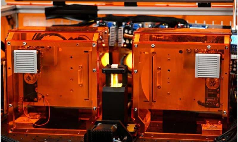
This pioneering spirit is perfectly reflected in the printer, an early tissue printing prototype, whose sheer orange acrylic casing reveals an inside of wires, projectors, copper coils, and tiny digital displays.
While it may seem speculative for now, the detailed and lightning-fast printing of living tissue in a suitcase-sized, orange 3D printer is completely real.
“Our research is a necessary first step for the future of tissue engineering. Right now, it can print more physiologically relevant 3D environments for cell culture, and in the long term, it could help make 3D-printed organs a reality,” says Dias Castilho.
Tissue printing with light
At the heart of the machine sits a tiny cuvette containing a fluid that transforms into a solid as if by magic. But instead of waving a magic wand, Lena Stoecker, who is a Ph.D. of Dias Castilho’s brand-new Biomaterials Engineering and Biofabrication group, projects beams of light onto liquids to conjure up viable cell-laden geometries.
Stoecker has successfully adapted a novel 3D printing technique called Xolography to print biomaterials. While demonstrating the printer by putting a cuvette with a liquid inside, Stoecker explains what drew her to 3D printing tissues: “I first encountered 3D printing as a student assistant during my studies of mechanical engineering and business administration. We employed 3D printing mainly for prototyping and tooling for small series production, and I was fascinated by the technology’s possibility to realize (almost) any idea.”
Biomedical challenges
It is no surprise that Stoecker gravitated towards tissue engineering, as it is by nature a multidisciplinary field combining the expertise of molecular biologists, engineers and designers.
The biggest trifold challenge facing tissue engineers everywhere is to create viable 3D tissues that closely resemble the natural environment of cells, to create them fast, and to do it precisely. This is the holy grail.
“There was a big hype around 3D printing for biomedical engineering, but technologies failed to meet the high expectations,” Stoecker explains. “My dream for Xolography would be to develop into a technology that is actually able to create tissue and organ models to study disease and develop cures.”
A technique from the field of design
Xolography is a groundbreaking fusion of engineering, physics and chemistry, where light is used to 3D print liquid polymers. It harnesses the power of intersecting light beams of distinct wavelengths within a light-reactive fluid. As light rays converge, they turn the fluid into a detailed, solid 3D object the size of a gummi bear in under a minute.
The technology was developed by German chemist Stefan Hecht and physicist Martin Regehly, who further adapted it for diverse applications in their spin-off business Xolo. Four years ago, Hecht mused about Xolography potentially being used for generating complex biological structures.
Dias Castilho explains, “Four years ago, Xolo was looking to advance its technology into biomedical applications, while my team was searching for a disruptive technology that could potentially offer high resolution, fast manufacturing speeds, and scalability—so it’s a perfect marriage.”
Today, the TU/e-researchers at the Biomaterials Engineering and Biofabrication group made printing tissue with light a reality. Hecht and Regehly follow the findings of the research group with interest, as they are the first scientists to use this technology to print living materials in the world.
That did not happen overnight, as the researchers had to overcome some additional challenges to adapt Xolography to printing living tissue.
“The materials used must be biocompatible, for one. Besides the hydrogels we were developing for the process, we found that the photoinitiator system itself was not very cell-friendly and had to be replaced. In close collaboration with the company, we developed and optimized the material formulations to ensure they are safe for biomedical applications,” says Dias Castilho.