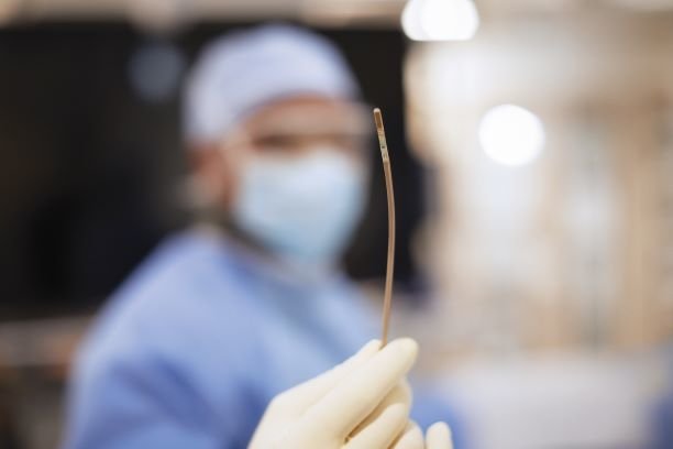
Philips’ 3D intracardiac echocardiography catheter is ready for its close-up—its extreme, in vivo close-up, that is.
A little less than a year after securing 510(k) clearance from the FDA, the VeriSight Pro treated its first patient outside clinical trials. A surgical team at the Mayo Clinic used the catheter, which captures echocardiograms to map the inside of the heart, in a left atrial appendage occlusion procedure.
In that procedure, a sac embedded in the wall of the left atrium known as the left atrial appendage is sealed off to keep blood clots from making their way to the heart itself. It typically requires an ultrasound probe to be passed through a sedated patient’s throat and into the esophagus to provide live imaging.
With the VeriSight Pro, however, that same imaging technology has been shrunk down to fit on the end of a three-millimeter catheter that is inserted into an artery at the neck, arm or groin. The patient doesn’t need to be sedated, which eliminates the risks and restrictions that come with general anesthesia.
Once the catheter reaches the patient’s heart, it uses intracardiac echocardiography technology to provide real-time 2D and 3D imaging data. Those images are transmitted to monitors on Philips’ EPIQ CVx cardiac ultrasound system to help guide cardiologists through their procedures.
The VeriSight Pro’s real-time imaging tools include the ability to view two 3D scan planes at the same time, allowing, for example, a cardiologist to simultaneously assess both axes of the left atrial appendage to determine which device should be used to close it.
The system also provides high-quality 2D images of the structures, valves and blood flow inside the heart. Because of the way the camera is positioned on the tip of the catheter, physicians can capture scans from multiple views and angles throughout the heart’s interior without needing to manually reposition the catheter.
The VeriSight Pro is designed to be used in minimally invasive procedures in structural heart disease and electrophysiology. The device is currently available only on a limited basis in the U.S.
The “next-generation” technology can transform some complicated procedures, said Mohamad Adnan Alkhouli, the Mayo Clinic interventional cardiologist who performed the first procedure with the VeriSight Pro. For one thing, it makes a left atrial appendage occlusion accessible to many patients who aren’t good candidates for general anesthesia, he said.
Alkhouli also noted that the catheter’s “excellent imaging of the tricuspid valve” also makes it a more effective treatment tool for tricuspid regurgitation, in which a malfunctioning valve allows blood to flow back into the heart’s upper right chamber.
Philips has been on a roll in the image-guided therapy department in the last several months. In April, it nabbed FDA clearance for the SmartCT imaging software, which integrates with the recently relaunched Azurion platform to guide physicians through capturing and analyzing 3D scans collected through angiography, neurology, soft-tissue imaging and guidewire navigation.
The same month, Philips reported massive gains in its diagnosis and treatment business for the first quarter of 2021. With an 11% increase in orders during the period, that segment brought in an impressive $2.24 billion in sales, leading Philips to adopt more optimistic projections for its post-COVID recovery.

