
Autism spectrum disorder (ASD), commonly referred to as autism, is a neurodevelopmental condition characterized by differences in social interactions, communication, behavior and the processing of sensory stimuli. Notably, the experiences, aptitudes and needs of autistic people can vary significantly.
Some neuroscientists have been exploring the possibility that this well-documented diversity partly reflects differences in the brain’s organization and underlying neurobiology. However, so far only a few studies have been able to link differences in autistic behavior to specific neurobiological processes.
Researchers at the Italian Institute of Technology’s Center for Neuroscience and Cognitive Systems (CNCS@UNITN) and the Child Mind Institute in New York carried out a study aimed at better delineating the brain connectivity patterns associated with autism.
Their findings, published in Nature Neuroscience, led to the identification of two distinct autism subtypes characterized by distinct connectivity patterns.
“This study was inspired by a certain frustration with how neuroimaging findings in autism have often been interpreted,” Alessandro Gozzi, senior author of the paper, told Medical Xpress.
“Autism is extremely heterogeneous clinically, and for many years imaging studies have also reported heterogeneous and sometimes apparently conflicting findings: some studies found reduced functional connectivity, others found increased connectivity, and others found more complex patterns.
“This led to a long debate about what this variability means. In many cases, it was treated as noise, a manifestation of the reproducibility crisis in neuroscience, or as a problem to be averaged away.”
In collaboration with Adriana Di Martino and her colleagues at the Child Mind Institute, Gozzi and his team at IIT set out to test a new hypothesis. Specifically, they hypothesized that widely reported differences in the brain connectivity patterns of autistic individuals are not random “noise,” but they are instead biologically meaningful.
“In other words, we wanted to determine whether different patterns of brain connectivity in autism reflect different underlying biological mechanisms,” said Gozzi.
“To test this idea rigorously, we designed a cross-species study. We started from mouse models, because in mice we can study autism-relevant genetic and biological perturbations under controlled experimental conditions, and we can more directly interrogate causal mechanisms.”
Studying the connectivity patterns linked to autism
As part of their study, Gozzi and his colleagues studied the brain connectivity patterns associated with 20 different mouse models of autism, as well as those captured in human patients. These are mice that are genetically engineered in different ways that prompt them to exhibit behaviors like those observed in autistic people.
First, the researchers tried to determine whether the highly varied connectivity patterns that emerged across different mouse models of autism could be linked to specific molecular and cellular processes. Subsequently, they tried to determine whether the same patterns could also be observed in brain imaging data collected from autistic people.
“Our broader goal was to turn what has often been seen as a limitation of autism imaging—its variability—into a mechanistic clue,” said Gozzi. “Rather than asking whether the autistic brain is simply more connected or less connected, we asked whether there are distinct connectivity subtypes that point to different forms of underlying biology.”
To capture the connections between different brain regions in both mice and humans, the team used resting-state functional MRI (fMRI). This is an imaging technique that records brain activity that spontaneously emerges when humans and animals are awake but not engaged in any task, by tracking the flow of blood in the brain.
“In imaging neuroscience, we quantify this communication using a measure called functional connectivity,” explained Gozzi. “Put simply, if the activity of two brain regions fluctuates together over time, we infer that these regions are functionally connected.”
“Our approach had three main steps. First, we measured functional connectivity across a large panel of mouse models relevant to autism, each carrying different autism-associated genetic or biological perturbations. Second, we asked whether these models naturally grouped into distinct connectivity patterns, and whether those patterns could be linked to different molecular or cellular mechanisms.”
The final part of the team’s study was aimed at determining whether the connectivity subtypes observed in mice, or corresponding subtypes, were also present in humans. To do this, Gozzi closely collaborated with Di Martino and her colleagues at the Child Mind Institute, who collected brain scans from autistic and non-autistic children.
“The key idea was to use the mouse data as a biological “Rosetta Stone: if a given connectivity pattern in mice is associated with synaptic biology, or with immune-related mechanisms, we can then look for similar patterns in human brain scans,” said Gozzi. “In this way, the animal models helped us interpret human imaging heterogeneity in mechanistic terms, rather than only describing it statistically.”
Two different autism connectivity subtypes
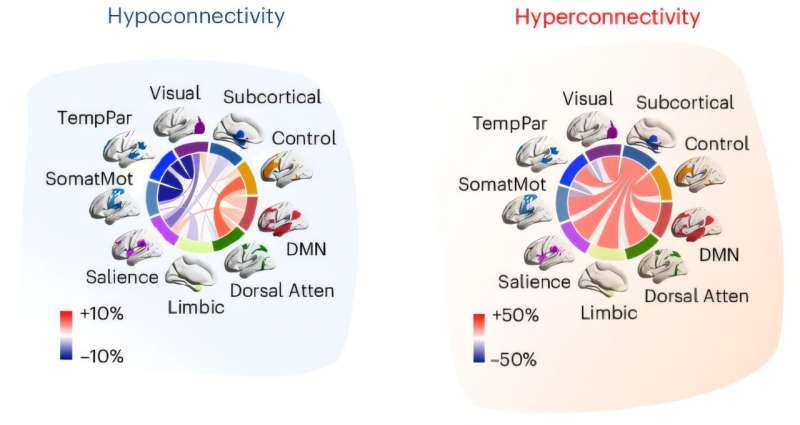
The analyses carried out by Gozzi, Di Martino and their colleagues ultimately led to the identification of two different patterns in functional connectivity that were observed both in mouse models of autism and humans.
The first of these patterns was characterized by a reduced communication between brain regions (i.e., hypoconnectivity), while the other entailed an increased communication between brain regions (i.e., hyperconnectivity).
“Importantly, these patterns did not simply entail ‘more’ or ‘less’ connectivity everywhere, but organized brain-wide patterns that could be linked to distinct biology,” said Gozzi.
“This suggests that at least part of autism heterogeneity can be parsed into biologically meaningful brain-connectivity subtypes. The hypoconnected subtype was linked to synaptic pathways, suggesting altered communication between neurons.
“By contrast, the hyperconnected subtype was associated with immune-related pathways and with alterations in gene regulation, suggesting that neuroimmune mechanisms and dysregulated transcriptional programs may contribute to a different form of circuit dysfunction.”
The team’s observations could improve the neuroscientific understanding of autism. If they are validated in further studies, they could eventually also inform the development of new support tools and practices that account for differences between individual patients.
“Two individuals may both receive an autism diagnosis, and may even show overlapping behavioral features, but the brain and molecular mechanisms contributing to their condition could be quite different,” said Gozzi.
“That distinction matters if we want to move toward more precise and personalized interventions. I would emphasize that this is not yet a clinical diagnostic tool. However, it provides a framework for future precision psychiatry in autism.
“Instead of asking only whether the autistic brain is ‘more connected’ or ‘less connected,’ we can begin to ask which circuit-level subtype is present, what biology it reflects, and whether different subtypes might respond differently to interventions.”
Directions for future research
The findings of this study could soon inspire more studies exploring the functional connectivity patterns associated with ASD. Meanwhile, Gozzi and his colleagues are planning further research aimed at understanding what experiences and behavioral differences might be associated with the two connectivity subtypes that they uncovered.
“Together with Di Martino and her colleagues, we want to apply this approach to human datasets that include not only high-quality brain imaging, but also deep phenotyping: detailed information about cognition, sensory symptoms, development, adaptive functioning, clinical history, genetics, and other biological measures,” explained Gozzi.
“This will be essential to understand how these brain-based subtypes relate to the real-world diversity of autism.”
As part of their next studies, Gozzi and his colleagues also hope to delineate the features of the autism subtypes they uncovered in greater detail. While they have so far identified two subtypes, they believe that there may be additional ones.
“If we can build richer and larger mouse imaging datasets, including more models and more biological perturbations, we may be able to partition this space more finely—perhaps identifying three, four, or more mechanistically distinct subtypes,” said Gozzi. “This will take years, but it is an important direction we are already working on.”
The researchers also hope to pin-point the physiological processes associated with hyperconnectivity and hypoconnectivity. They have already come up with a few hypotheses regarding these patterns’ underlying physiology, which they are currently testing experimentally.
“We are now trying to decode these fMRI signals in terms of neuronal activity, circuit dynamics, and excitation/inhibition balance,” added Gozzi. “Ultimately, we want to go beyond demonstrating that these connectivity patterns exist, also understanding what they imply for brain function and dysfunction.”