
Researchers from several Parisian institutions have worked together to develop a non-destructive approach to study how unicellular organisms respond to stress, focusing on cell-to-cell differences. Working together, the researchers combined custom fluorescence microscopy with machine learning. Together, they measured how individual algae cells protect their photosynthetic machinery from excess light. This revealed unexpected coordination between protective mechanisms that remains invisible when measuring cell populations in bulk.
The team was led by Aliénor Lahlou from École Normale Supérieure—PSL University, in collaboration with Sony CSL—Paris and the Institut de Biologie Physico-Chimique. The research results, published in New Phytologist, demonstrate how single-cell analysis can uncover biological strategies that traditional methods miss, which could help improve hypothetical models and select individual traits relevant for biotechnologies.
A new window into photosynthetic stress responses
Most knowledge about photosynthesis comes from measuring whole plants or large populations of microalgae. While such studies have provided fundamental insights, they average out the natural variation between individual cells—variation that can reveal how organisms orchestrate their responses to environmental stress.
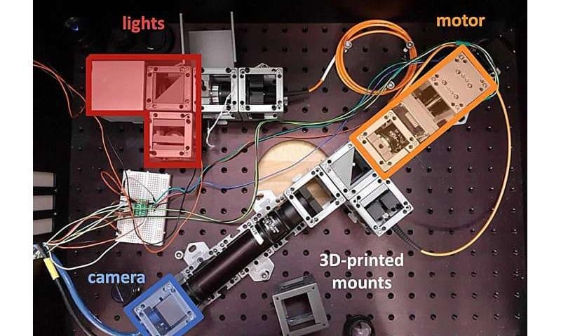
The research team developed an automated fluorescence microscope capable of simultaneously tracking hundreds of individual Chlamydomonas reinhardtii cells as they respond to controlled light exposure. The system measures chlorophyll fluorescence—a widely used indicator of photosynthetic status and stress—with sufficient sensitivity to detect three distinct protective mechanisms called non-photochemical quenching (NPQ) components.
The technical challenge lies in separating these three NPQ components, which occur simultaneously over timescales ranging from seconds to hours, and create complex, overlapping fluorescence signatures. The team’s solution leverages a key biological insight: By using genetic mutants and specific growth conditions, they created reference populations of algae expressing only one NPQ component at a time. These “training populations” taught machine learning algorithms—specifically dictionary learning and linear discriminant analysis—to recognize the characteristic fluorescence patterns of each component.
The result is a three-dimensional reference space where any new fluorescence temporal traces can be projected and assigned three scores—one for each NPQ component—without requiring complex mathematical assumptions.
Hidden coordination emerges from individual variation
Even among synchronized, genetically identical algae, individual cells showed substantial differences in their NPQ responses—with variation coefficients of approximately 40% for high-energy quenching (qE) and 20% for state transitions (qT). The team demonstrated this variation reflects real biological differences rather than experimental noise by measuring the same cells repeatedly.
More surprisingly, they discovered a strong correlation between qE and qT at the single-cell level: Cells developing stronger qE showed weaker qT, and vice versa. This trade-off persisted across populations exposed to different durations of high-light treatment, suggesting active cellular coordination between the two mechanisms—a relationship completely masked in bulk measurements where individual responses are averaged together.
Intriguingly, a different wild-type strain showed similar trends at the population level but lacked the individual-cell correlation. Some cells in this strain maintained both high qE and high qT simultaneously—a phenotype never observed in the first strain. This demonstrates that genetically distinct individuals can orchestrate the same protective mechanisms very differently, even when their population averages look identical.
Broader applications and future directions
Because the machine learning framework relies solely on signal dynamics without requiring prior knowledge of specific temporal patterns, the researchers noted it should be adaptable to other photosynthetic organisms and stress conditions—including nutrient deficiency or extreme temperatures—provided these stresses trigger detectable fluorescence changes.
However, the approach has important limitations. It requires building a training dataset with reference populations expressing isolated components, which restricts application to organisms where the necessary genetic tools and biological knowledge exist. The team experienced this challenge when including photoinhibition (qI) in their analysis, since the underlying processes are less well-controlled than for qE and qT.
Additionally, the method assumes that complex cellular responses can be satisfactorily reconstructed from combinations of elementary responses—a hypothesis that must be validated for each new application through reconstruction quality metrics.
The researchers see particular promise in integrating their non-invasive fluorescence approach with rapidly advancing flow cytometry, microfluidics, and single-cell omics methods. This could enable correlations between cell morphology, genotypes, and photosynthetic traits, facilitating applications such as varietal selection, directed evolution, screening after mutagenesis, or isolation of species from natural samples.
Combining with single-cell omics as a final step—since that analysis is destructive—could identify the genetic determinants underlying the observed heterogeneity in more detail.