
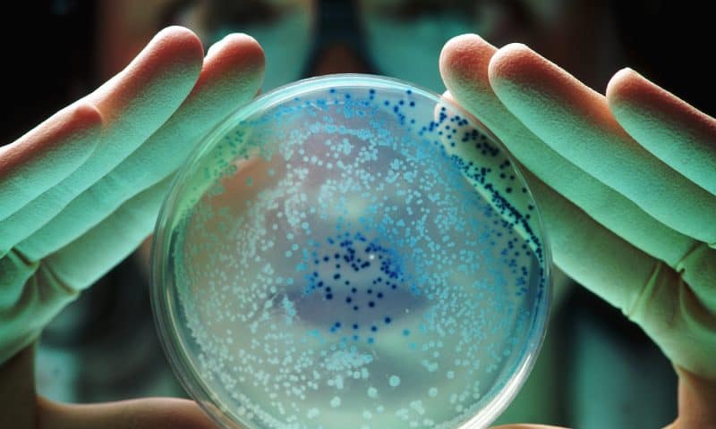
Scientists from Cornell University, University of California, San Francisco (UCSF), and elsewhere have found that a surplus of membrane proteins may help bacteria survive antibiotic exposure. These proteins are part of a shuttling mechanism that bacteria use to pump out a wide spectrum of antibiotics along with other physiological substrates from the cell. The researchers are now focused on using chemical and mechanical manipulations to disrupt the process so that antibiotics can be more effective.
Details of the study were published in Cell Reports Physical Science in a paper titled, “Transporter excess and clustering facilitate adaptor-protein shuttling for bacterial efflux.”
In it, the researchers describe how an imbalance in the three-part protein complex—MacAB-TolC—helps gram-negative bacteria resist antibiotics. The MacAB-TolC complex, known as a multidrug efflux pump, spans the cell’s inner and outer membranes, as well as the periplasm that connects them. Each protein occupies a different location: TolC on the outer membrane, MacB on the inner membrane, and MacA in the periplasm, although it is anchored on the inner membrane. This protein complex forms a conduit that drains out antibiotics as well as virulence factors produced by the bacterial cell.
The three proteins need to assemble in a specific stoichiometry to pump out toxins. Two MacB proteins assemble with six MacA proteins, then three TolC proteins. Scientists understand this ratio well. What’s not been clear is how molecules in the periplasm enter the channel that runs through the complex and through which substrates they get pumped, once the structure is assembled.
“You basically have these extra Bs that don’t have A partners to assemble. And of course, the cell does not do this for no reason,” said Peng Chen, PhD, study lead and a professor of chemistry at Cornell University. “We found out a good reason is that when you have this extra B, because it does not have A associated with it, it naturally has an opening for the substrate to go in. So once the substrate can bind to the extra B, some of the As that are initially associated with B can migrate over to assemble. And once it’s assembled, they can pump the substrate out.”
As part of the study, the researchers tested whether the mechanism could be interrupted. They used a microfluidic device developed at UCSF that applies mechanical stress to change bacteria’s toxin resistance. They found that squeezing E. coli through the device deformed the cell enough to disrupt the assembled complex and prevent it from resisting antibiotics.
The scientists believe that the behavior observed in E. coli likely exists in other systems. “This imbalance of protein stoichiometry must exist for many types of protein complexes. But how does a cell utilize this imbalance?” Chen said. “Now we have one example that shows this particular imbalance might be, functionally, very relevant. So anytime that we study protein complexes in the cell, we always want to measure the relative amount in the entire cell versus their relative amount in a particular complex. Do they actually match?”